Miniature version of CERN detector to help treat brain tumors
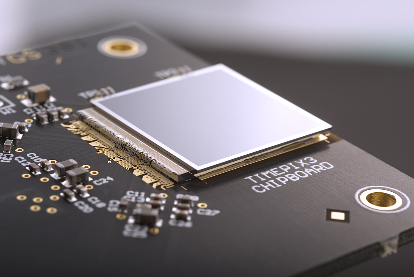
The Timepix3 chip developed at CERN to detect atomic particles is now finding applications in medical imaging and can help direct radiation treatment.

A new imaging device built by miniaturizing the particle detector technology used by the experiments at CERN is now being used to aid brain tumor treatments. Czech company ADVACAM developed the device, and it is being tested through institutional collaboration in Germany, according to a press release.
Setting up humungous facilities like CERN that carry out a handful of experiments yearly is an expenditure that countries can avoid during testing economic times. Even as the large-scale benefits of this research can take years and even decades to realize, science also leads to smaller wins that can make a world of difference in other fields.
The imaging device built by ADVACAM is one such example. Built on the original design of the detector that spots particles being dispersed in the accelerator, the Timepix3 pixel detector can help direct brain tumor treatments accurately.
How brain tumors are treated
Tumors can be treated with powerful radiation. This involves using ion beams that travel closer to the speed of light and through tissues. The difficult part, however, is directing the radiation only to cancerous cells and not to healthy ones.
This is achieved by moving the ion beams in an arc where the tumor is at the center. This allows maximum radiation to be directed to the cancerous cells. In contrast, only a small percentage of healthy cells are exposed.
While this works for most other tissues, the approach is tricky for tumors located inside the brain. Even the slightest overexposure of healthy cells to powerful radiation risks outcomes such as damage to optic nerves or loss of memory.
To minimize these risks, a computerized tomography (CT) scan is usually used to locate the tumor and guide the radiation therapy accurately. However, the scan can be misleading since the brain position may have moved inside the skull during this duration.
Accurate imaging with a new detector
The team at ADVACAM used the accurate detection capabilities of detectors at CERN to build a small version to track secondary radiation from the ion beam directed at tumor cells. Using this radiation, the researchers created a tissue map to confirm whether the beam was being directed accurately.
“Our cameras can register every charged particle of secondary radiation emitted from the patient’s body,” said Lukáš Marek, who was involved in developing the detector at ADVACAM. “It’s like watching balls scattered by a billiard shot. If the balls bounce as expected according to the CT image, we can be sure we are targeting correctly. Otherwise, it’s clear that the ‘map’ no longer applies.”
The approach is currently being trialed by scientists from the German National Center for Tumor Diseases (NCT), the German Cancer Research Center (DKFZ), and the Heidelberg Ion Beam Therapy Center (HIT) at Heidelberg University Hospital.
The treatment must be stopped when the clinicians determine that the radiation targeting is inaccurate. In the future, ADVACAM intends to allow corrections to happen in real-time so that patient’s treatment is not delayed, the press release added.
“When we started developing pixel detectors for the LHC we had one target in mind – to detect and image each particle interaction and thereby help physicists to unravel the secrets of Nature at high energies.” said Michael Campbell, a spokesperson of the Medipix Collaborations who developed the detectors for ADVACAM. “The Timepix detectors take the same technology to new fields that were completely unforeseen at the beginning. This application is a brilliant example of that.”
 SHOW COMMENT ()


