Article

Shoot Development in Members of an Ancient Aquatic Angiosperm Lineage, Ceratophyllaceae: A New Interpretation Facilitates Comparisons with Chloranthaceae

Faculty of Biology, M.V. Lomonosov Moscow State University, 119234 Moscow, Russia
*
Author to whom correspondence should be addressed.
Symmetry 2022, 14(7), 1288; https://doi.org/10.3390/sym14071288
Submission received: 4 April 2022 / Revised: 12 June 2022 / Accepted: 17 June 2022 / Published: 21 June 2022
(This article belongs to the Section Life Sciences)

Abstract

:
Ceratophyllum is an ancient and phylogenetically isolated angiosperm lineage. Comparisons between Ceratophyllum and other angiosperms are hampered by uncertainty in inferring organ homologies in this genus of specialized aquatics. Interpretation of shoot morphology is especially problematic in Ceratophyllum. Each node has several leaf-like appendages interpreted as verticillate leaves, modified parts of one and the same leaf or parts of two leaves under decussate phyllotaxis. Vegetative branches are axillary, but reproductive units (interpreted as flowers or inflorescences) are commonly viewed as developing from collateral accessory buds. We studied shoot development in Ceratophyllum submersum, C. tanaiticum, and C. demersum using scanning electron microscopy to clarify shoot morphology and branching patterns. Our data support the idea that the phyllotaxis is essentially decussate with appendages of stipular origin resembling leaf blades. We conclude that a leaf axil of Ceratophyllum possesses a complex of two serial buds, the lower one producing a vegetative branch and the upper one developing a reproductive unit. The reproductive unit is congenitally displaced to the subsequent node, a phenomenon known as concaulescence. Either member of the serial bud complex may be absent. There is a theory based on a synthesis of molecular and morphological data that Chloranthaceae are the closest extant relatives of Ceratophyllum. Serial buds and concaulescence are known in Hedyosmum (Chloranthaceae). Our new interpretation facilitates morphological comparisons between Hedyosmum and Ceratophyllum.

1. Introduction

The occurrence of axillary branching is among key morphological characteristics of seed plants [1,2]. The term axillary branching implies that each lateral axis (including vegetative branches as well as lateral strobili, flowers, or inflorescences) develops immediately above (i.e., in axil) of certain phyllome, which is called a subtending phyllome. The subtending phyllome may represent a foliage leaf, cataphyll, or bladeless phyllome of reproductive region (bract). Free-sporing land plants often possess radically different patterns of shoot branching [3]. For example, lycophytes have terminal branching (dichotomy), horsetails and heterosporous ferns possess lateral branches in stem nodes side-by-side with leaves, and some homosporous leptosporangiate ferns develop branches on the abaxial side of their leaf bases.
Despite the importance of axillary branching as seed-plant character, deviations from this typical pattern are well-known in some scattered angiosperms and gymnosperms. Botanists tend to exploit morphological concepts explaining these deviations. This practice allows maintaining the idea of a common seed plant body plan. For example, the concept of accessory buds (which are either collateral or serial) is proposed to describe the occurrence of more than one bud per leaf axil [4]. The concept of metatopic displacement describes situations when a branch is attached to the main axis well above the putative subtending phyllome (concaulescence) or a branch and its subtending phyllome have a common base (recaulescence) [5]. In certain instances, an idea of evolutionary reduction of subtending phyllomes is useful [6,7,8,9]. For example, most Brassicaceae—including the wild type Arabidopsis thaliana—possess no bracts subtending lateral flowers, but it is commonly accepted that such bracts were present in ancestors and became lost in the course of evolution. In a few seed plants, however, even use of ad hoc hypotheses (such as metatopic displacement or bract reduction) reportedly cannot defend the occurrence of axillary branching and their ramification patterns could be fundamentally different from those in the vast majority of angiosperms and gymnosperms. For example, dichotomous branching has been described in cycads [10]. Unfortunately, the scattered seed plant taxa with reportedly dichotomous branching are problematic for SEM-based developmental studies using ample material. One cannot expect getting fixed samples of numerous vegetative shoot apices of any cycad captured at early stages of branching.
There are two ancient angiosperm lineages—Nymphaeales and Ceratophyllales—that are conspicuous in the occurrence of deviations from axillary branching, especially with respect to flower arrangement [8,11,12,13,14,15,16,17,18,19,20,21,22,23,24,25]. Both lineages are aquatic and not sister to each other. While the phylogenetic placement of Nymphaeales in the basal angiosperm grade is currently well-established, the relationships of Ceratophyllales are incompletely resolved [26,27,28]. Ceratophyllales, with the only extant family Ceratophyllaceae, the only extant genus Ceratophyllum (sometimes two genera are recognized [29]), and six species [30], is the smallest of the five primary lineages of mesangiosperms. Other mesangiosperm clades are eudicots, monocots, magnoliids, and Chloranthaceae. Recent molecular phylogenetic and phylogenomic data tend to place Ceratophyllum as sister to eudicots [27,31], but analyses that use data on morphology of extant and fossil angiosperms [8,32,33,34], as well as some molecular-based studies [35,36,37,38], support a hypothesis that the closest relatives of Ceratophyllum living today are Chloranthaceae.
All species of Ceratophyllum are specialized aquatics with underwater pollination [25,39,40,41,42,43,44,45,46,47]. They lack any traces of roots. At first glance, they are characterized by verticillate phyllotaxis with numerous leaves per node (Figure 1). These ‘leaves’ (called ‘leafy appendages’ below) are dichotomously divided 1–4 times into narrow segments. More detailed observations have challenged the idea of verticillate phyllotaxis. Schaeppi [48] summarized evidence for the occurrence of decussate phyllotaxis in Ceratophyllum. Under this interpretation, leafy appendages of each node belong to two strongly dissected opposite leaves. Further refinement of this interpretation is provided by Iwamoto et al. [25] who interpreted all but central leafy appendage of the opposite leaves as multiplied stipules (stipule-like appendages). On the other hand, Raynal-Roques [22] developed a theory that all appendages of each whorl belong to one and the same dissected leaf.
Homologies of structures bearing stamens and carpels in Ceratophyllum are problematic [25,39,40,42,43,44,45,46,47,49,50,51]. Therefore, we use a neutral term ‘reproductive unit’ [50]. Each reproductive unit has a whorl of involucral appendages. These are similar to leafy appendages, but undivided. In male reproductive units, the involucre is followed by stamens arranged along the short axis of the unit. Each female reproductive unit has an uniovulate gynoecium inside the involucre. The reproductive units of Ceratophyllum were often interpreted as unisexual flowers with a perianth [41,52,53]. However, the idea of tepal homologies of involucral phyllomes is problematic because of the occasional occurrence of additional reproductive unit(s) inside the involucre [40,43,47]. According to Endress [51] and Endress and Doyle [47], each stamen of Ceratophyllum should be viewed as an individual male flower possessing no subtending bract.
Earlier studies of Ceratophyllum demersum and C. submersum revealed that patterns of arrangement of lateral axes (vegetative branches and reproductive units) follow strong regularities [22,23,24,25,39,48]. The lateral axes form orthostichies along the main axis. The most complete set of orthostichies is four (Figure 2A), but two (Figure 2B) or one of them are often missing or incomplete in real plants. The more complete orthostichies will be called below leading orthostichies. When two leading orthostichies are present, they are in adjacent positions and the angle between their radii is estimated at about 80° [22] or 70–90° [24]. The shoot side bearing leading orthostichies is oriented towards the water surface. Each vegetative branch normally develops in a radius of a leafy appendage of a main axis node [23,24,25,39,48]. Thus, the branch position is interpreted as axillary and the subtending appendage is viewed as a median leaflet or leaf blade.
Under the decussate interpretation of phyllotaxis, a maximum of two vegetative branches per node can be expected (Figure 2A). When all branches belong to two adjacent orthostichies, one of two branches is systematically suppressed in each node on one side of the dorsiventral shoot (Figure 2B), a phenomenon known as sectorial anisoclady [24]. Deviations from equal spacing (90°) of the four orthostichies observed in Ceratophyllum can be regarded as one of manifestations of shoot dorsiventrality. Similar phenomena are observed in some other plants with dorsiventral shoots and decussate phyllotaxis (e.g., Selaginella).
The occurrence of distichous phyllotaxis (Figure 2C,D) was postulated in Ceratophyllum using material with all vegetative branches belonging to the two leading orthostichies [22]. Under this interpretation, divergence angles between adjacent leaves are strongly unequal along the shoot axis: 80°–280°–80°–280°–80°–280°, a phenomenon known in some angiosperms with a dorsiventral kind of distichous phyllotaxis, or pendulum symmetry [54,55,56].
The reproductive units always develop between the radii of the leafy appendages of the main axis [23,24,25,39,40,43,48]. Assuming their positions relative to vegetative branches (Figure 2A,B), the reproductive units can be interpreted as extra-axillary structures. Their positions exactly fit the expected boundaries between the two leaves of a node under the decussate interpretation (Figure 2A,B). Under the hypothesis of dorsiventral distichy, the reproductive units can be interpreted as developing from accessory—more precisely, collateral—buds situated side-by-side of the principal bud, developing a vegetative branch [22]. Raynal-Roques [22] found occasional occurrence of four (male) reproductive units per node with two units closer to the vegetative bud being larger than the other two units (Figure 2C), a condition that can be accommodated by the idea of collateral buds [22]. Iwamoto et al. [25] followed the hypothesis of decussate phyllotaxis and at the same time accepted the origin of reproductive units from collateral buds. In their view, each of the two opposite leaves may have a maximum of two accessory buds developing reproductive units; the condition with four units takes place when both leaves develop two accessory buds. The condition when all reproductive units belong to the same orthostichies as vegetative branches can be acquired via suppression of one accessory bud per leaf and certain lateral shift of the remaining accessory bud (Figure 2E).
Earlier studies of shoot architecture and branching patterns in Ceratophyllum mainly covered two species, C. demersum and C. submersum. The present paper provides comparative SEM-based developmental data on three species of the genus (C. submersum, C. tanaiticum, and C. demersum) representing different clades revealed using molecular phylogenetics [30]. The aims of our study are further refinement of morphological interpretation of Ceratophyllum, analysis of developmental correlations and search of potential interspecific differences. Robust morphological interpretation of plant structure is essential for comparisons of Ceratophyllum with members of other angiosperm lineages and for character scoring in morphological datasets. We believe that our study provides new insights important for resolving the enigma of mesangiosperm radiation.

2. Materials and Methods

Whole plants and young shoot tips of Ceratophyllum demersum, C. submersum, and C. tanaiticum were fixed in 70% ethanol. Samples of C. demersum were collected from a pond in Zvenigorod Biological Station of Moscow State University (Odintsovsky distr., Moscow Prov.; voucher: Sokoloff & Remizowa s.n., 2016, MW). Samples of C. tanaiticum and C. submersum collected in Khoper State Nature Reserve, Voronezh prov., Russia were kindly provided by E.V. Pechenyuk (vouchers: Pechenyuk 16, 2012; Pechenyuk 32, 2017, MW). About 15 shoots of each species were studied. For scanning electron microscopy (SEM), material was dissected in 70% ethanol and transferred to 100% acetone using the following series: 96% ethanol (twice for 30 min), 96% ethanol: 100% acetone (1:1 v/v, 30 min), 100% acetone (three times for 30 min). The material was critical-point dried using a Hitachi HCP-2 critical-point dryer (Hitachi, Tokyo, Japan), then coated with gold and palladium using a Eiko IB-3 ion-coater (Tokyo, Japan) and observed using a CamScan S-2 (Cambridge Instruments, London, UK) at the Laboratory of Electron Microscopy at the Biological Faculty of Moscow University.
Descriptive terminology used in the paper is summarized in Box 1.
Box 1. Descriptive terminology used in the paper
Accessory buds: additional buds developing in a leaf axil.
Axillary branch: a branch that has a subtending phyllome.
Bud: incipient shoot.
Collateral buds: a group of one principal and one or more accessory buds situated in the axil of the subtending phyllome and attached at the same level in a horizontal plane.
Concaulescence: attachment of an axillary bud well above its subtending phyllome along the stem of the mother shoot.
Decussate phyllotaxis: phyllotaxis with two leaves per node; leaves of neighboring nodes are attached at regularly alternating radii; all leaves of a shoot form four orthostichies.
Distichous phyllotaxis: phyllotaxis with one leaf per node; all leaves of a shoot are arranged along two orthostichies.
Internode: a portion of stem between two neighboring nodes.
Involucral appendage: an interpretation-neutral term used here to name individual leaf-like structures belonging to reproductive units. A whorl of involucral appendages surrounds a pistil or stamen(s) of each reproductive unit. Depending on interpretation, each leafy appendage represents an entire phyllome or several appendages collectively form a phyllome.
Leafy appendage: an interpretation-neutral term used here to name individual leaf-like structures developing in stem nodes in Ceratophyllum. Depending on interpretation, each leafy appendage represents an entire phyllome or several appendages collectively form a phyllome.
Node: a short portion of stem where a leaf (or leaves) is attached.
Orthostichy: a virtual vertical line of stem surface along which a set of leaves is attached; in other words, all leaves belonging to an orthostichy are attached to the stem at the same radius.
Phyllome: a general term for typical leaves and all their homologs (for example, foliage leaves, bud scales, bracts, sepals).
Phyllotaxis: pattern of arrangement of leaves along the stem.
Reproductive unit (RU): compact part of plant body bearing stamen(s) and/or pistil(s). This interpretation-neutral term can be used when precise recognition of flowers is problematic.
Serial buds: a group of one principal and one or more accessory buds situated in the axil of the subtending phyllome and arranged along a vertical line
Shoot: stem with leaves and lateral buds.
Stipules: paired outgrowths at the leaf base.
Subtending leaf: a leaf in which axil (i.e., just above its insertion level) a lateral bud, shoot, or flower is situated.
Verticillate phyllotaxis: phyllotaxis with more than two (often many) leaves per node.

3. Results

3.1. Ceratophyllum submersum

3.1.1. Young Primary Shoot of Seed Embryo and Initiation of Its Phyllotaxis

In this species, we investigated embryo morphology and pattern of organ arrangement in first nodes above the cotyledons (Figure 3). As in other species of the genus, the embryo has no primary root and the root pole ends blindly (Figure 3A). The two cotyledons are massive. There is a pair of much smaller organs situated almost at the level of the cotyledons in positions at the right angle to the cotyledonary plane (‘lf’ in Figure 3). These should be interpreted as the leaves of the first node above the cotyledons. With this interpretation, the cotyledons as well as the leaves of the node above them lack stipules. An interpretation that organs labeled ‘lf’ in Figure 3 are pairwise fused stipules of the cotyledons must be rejected, because it implies that next node leaves are situated in the same radii as the cotyledons. Starting from the second node above the cotyledons, each node has six organs (Figure 3B,C). These can be interpreted as two leaves, each bearing a blade and two stipules. The stipules resemble the blade in the size and shape. The phyllotaxis is decussate. The youngest stage that can be seen in Figure 3B (at the fifth node above the cotyledons) shows that the stipules and the blade develop from a common primordium. In subsequent stage, young leaves are three-lobed (the fourth node above the cotyledons in Figure 3C). Below we use a term ‘leafy appendage’ when we are not intending to distinguish between the morphologically similar stipular organs and leaf blades. Fully developed leafy appendages are divided dichotomously into filiform segments. Each segment has an apical mucilaginous gland (Figure 4A).

3.1.2. Initiation of Phyllotaxis in Lateral Shoots

Patterns of organ arrangement at the beginning of lateral shoots vary, but the first leaves (prophylls) can be inferred as occupying transverse positions. The lateral shoot in Figure 4A has six organs in the first node that can be interpreted as two leaf blades in transverse positions and four stipules. The second node continues the decussate phyllotaxis. A young second node leaf visible in Figure 4A has a blade that is larger than the two stipules. The lateral shoot in Figure 4B has more than six organs in the first node. The leafy appendages in transverse positions subtend next order lateral axes and can be plausibly interpreted as leaf blades. The occurrence of more than two stipules (or appendages derived from stipules) per leaf should be postulated in the first and subsequent nodes of the lateral shoot in Figure 4B. It can be easily seen that the third node leaf has a blade (la*) that subtends a next-order axis (vb*) and two pairs of stipules (la) on either side (Figure 4B).

3.1.3. Morphology of Mature Shoots

Shoots possess (6)8–12(13) leafy appendages a node. Mature leafy appendages of C. submersum are illustrated in our earlier paper (Figure 11C,D of [50]). The number of appendages often differs even among neighboring nodes (Figure 5). When the two adjacent nodes possess an equal number of the appendages, these are inserted in regularly alternating radii. The shoots possess certain nodes where, out of all angles between adjacent leafy appendages, one much exceeds the others. Such a wide gap always occurs directly above a vegetative branch of the previous node. An example is provided in Figure 6A (asterisk). Wide gaps between leafy appendages tend to occur every four nodes apart along the same orthostichy (Figure 5A: nodes 3, 7; Figure 5B: nodes 1, 5; Figure 5D: nodes 1, 5).
The maximum observed number of lateral axes per node is three (Figure 5). There are three types of lateral axes: vegetative branches, male reproductive units, and female reproductive units. The number of reproductive units in a node never exceeds two, and not more than two vegetative branches in a node have been observed. Thus, when three lateral axes are present, these are two reproductive units and a vegetative branch or two vegetative branches and a reproductive unit. Nodes with two lateral axes possess a vegetative branch and either a male or female reproductive unit. When only one lateral axis is present, this is either a vegetative branch or a female reproductive unit.
Patterns of arrangement of lateral axes observed in our sample of C. submersum are generally compatible with the ‘full general diagram’ of Ceratophyllum shoot (Figure 2A) assuming that some vegetative branches and reproductive units envisaged by the ‘full general diagram’ are missing. For example, in the shoot fragment illustrated in Figure 5D, only two orthostichies of lateral axes can be seen (as in Figure 2B); there is also a male reproductive unit in a position where a third orthostichy can be expected according to the ‘full general diagram’ (Figure 2A). The left-hand orthostichy in Figure 5D has reproductive units in nodes with odd numbers and vegetative branches in nodes with even numbers. The right-hand orthostichy has a reverse arrangement: reproductive units are in even nodes and vegetative branches are in odd nodes (Figure 5D). This is in full agreement with what is predicted by the general model for two adjacent orthostichies (Figure 2A,B). Notably, the pattern remains unchanged after nodes that possess no lateral axes in one (nodes 7, 8) or both (nodes 4, 5) orthostichies. Shoot fragments in Figure 5A,B show instances of minor deviations from the ‘full general diagram’. The deviations are highlighted by arrows. In both cases, a slight change in orthostichy position can be seen along the shoot length. Figure 5C shows an instance of a major deviation. This shoot can be thought as having two parts divided by an oblique dashed line. Patterns above and below the line as such are in agreement with the ‘full general diagram’, but transference of the pattern is broken at the dashed line. Therefore, in the left-hand orthostichy, vegetative branches are in odd nodes 1 and 7 above the line, but in an even Node 10 below the line.
In agreement with the ‘full general diagram’, all reproductive units are inserted in radii between two leafy appendages. Most vegetative branches are located in the axil of a leafy appendage, but some branches inserted in radii between two leafy appendages are also found (Figure 5B, Node 5; Figure 5D, nodes 2 and 6).

3.1.4. SEM-Based Description of Young Shoot Tips

Shoot apex is inclined to one side (Figure 6, Figure 7 and Figure 8A). The convex side of the shoot tip is directed towards the water surface. Developmental data show the occurrence of a leading orthostichy possessing regularly alternating vegetative branches (vb) and female reproductive units (fRU) in the following acropetal sequence: … fRU, vb, x, x, fRU, vb, x, x, fRU, vb, x, x …, where x are nodes bearing no lateral axes in the leading or any other orthostichy.
Vegetative branches of the leading orthostichy show a precocious initiation. A common primordium of vegetative branch and its subtending leafy appendage develops prior to other leafy appendages of the node (1vb in Figure 8A,B). When finally initiated, the leafy appendages are much smaller than the precocious branch of the same node (Node 1 in Figure 7; Node 4 in Figure 6). Positions of the appendages closest to the branch are shifted towards the proximal side of the shoot. These appendages are also delayed in development. The leafy appendage that subtends the branch (i.e., the putative leaf blade) is the last one to initiate as a distinct organ. As evidenced by analyses of definitive stages (Figure 5D), sometimes the subtending appendage fails to initiate at all. Reproductive units of the leading orthostichy initiate almost simultaneously with leafy appendages of their nodes and are located between—rather than above—the appendages (Figure 8A,B). Similarly to vegetative branches, the reproductive units show a precocious development. Already at the stage of two plastochrons from the apex, a reproductive unit much exceeds in size the leafy appendages of its node, though it is still smaller than just initiated vegetative branch above it (Figure 7A). Subsequent growth of female reproductive units is more rapid than those of vegetative branches and leafy appendages (Figure 6, Figure 7 and Figure 8A,B). Pistils become ready to pollination at only a short distance from the shoot apex (14fRU in Figure 7B).
Figure 7B illustrates a vegetative branch in a node right below a female reproductive unit (11vb), i.e., in a position where lateral axes are normally absent in C. submersum. Notably, this branch is much retarded in development or just recently initiated, as we never observed branches in such positions closer to the shoot apex. It is also slightly displaced from a position in the leading orthostichy by the large female reproductive unit (fRU).
Apart from the leading orthostichy, developmental data show the occurrence of some lateral axes in two other orthostichies adjacent to the leading one. These possess vegetative branches and male reproductive units. A node possessing a precocious vegetative branch in the leading orthostichy tends to possess a male reproductive unit in an adjacent orthostichy or in both adjacent orthostichies (nodes 4, 8 in Figure 6; nodes 5, 9 in Figure 8A,B). The reproductive units are larger than leafy appendages at early stages and are situated between—rather than above—the appendages (4RU in Figure 6B). The male reproductive units commence to anthesis at greater distances from the shoot apex than female units. They keep producing new stamens at the stage when the outermost stamens reach a definitive stage (Figure 8C–E). A node possessing a female reproductive unit in the leading orthostichy tends to possess a vegetative branch in an adjacent orthostichy or in both adjacent orthostichies (Node 5 in Figure 6; nodes 6, 10 in Figure 8A). Notably, these vegetative branches are delayed in development relative to those of the leading orthostichy.
Diagrams produced using definitive stages (Figure 5) show less precise patterns of arrangement of lateral axes than images taken from early developmental stages (Figure 6, Figure 7 and Figure 8). This may be related to late appearance of some lateral axes and possible occasional change of shoot orientation relative to water surface.

3.2. Ceratophyllum tanaiticum

With few exceptions discussed in subsequent paragraphs, our data on shoot morphology and development in C. tanaiticum (Figure 9, Figure 10, Figure 11, Figure 12, Figure 13, Figure 14 and Figure 15) resemble those on C. submersum. Patterns of arrangement of lateral axes observed in our sample of C. tanaiticum fit the ‘full general diagram’ of Ceratophyllum shoot (Figure 2A) assuming that some vegetative branches and reproductive units envisaged by the diagram are missing.
A common feature of C. submersum and C. tanaiticum is a retarded early growth of leafy appendages so that they are much shorter than most lateral axes in the nodes closest to the shoot apex, though some vegetative branches remain suppressed (Figure 9C,D). As in C. submersum, there is a tendency for the occurrence of rhythmic arrangement of lateral axes along the main axis, with a period of four nodes where lateral axes are missing in the third and fourth node of each period. For example, lateral axes are absent in nodes 5, 6; 9, 10; and 13, 14 in Figure 10 and in nodes 3, 4; 7, 8; and 11, 12 in Figure 11A. However, in the same way as in C. submersum, the rhythmic pattern is sometimes altered (Figure 13B). A vegetative branch normally develops in the axil of a leafy appendage (i.e., putative leaf blade). This subtending appendage is usually (Node 3 in Figure 10B, Node 4 in Figure 13B), though not always (Figure 13A) retarded in development relative to other leafy appendages of its node. Figure 14A (Node 2) shows two developmentally delayed leafy appendages in the sector of vegetative branch. None of them are in the branch radius. These patterns of developmental plasticity resemble those observed in C. submersum.
The shoot in Figure 11A has a leading orthostichy with regularly alternating female reproductive units and vegetative branches, as well as two adjacent orthostichies bearing only male reproductive units. The shoot in Figure 11B has two adjacent leading orthostichies, each bearing male and female reproductive units as well as vegetative branches. It is possible that the difference between the occurrence of one or two leading orthostichies is related to the position of shoot orthostichies relative to water surface (Figure 11C,D). The shoot in Figure 10 has a condition intermediate between the occurrence of one and two leading orthostichies: all vegetative branches belong to one and the same orthostichy along with female reproductive units, but a female unit is also found in another orthostichy along with male units. Figure 12 shows an instance of a change of leading orthostichy, possibly due to a shift of spatial orientation relative to the water surface. The leading orthostichy of the youngest part of the shoot is on the convex side of the tip. Apparently, this shoot has undergone a change in direction of tip curvature.
In our sample of C. tanaiticum, male reproductive units possess as few as one to three stamens (Figure 9C, Figure 10, Figure 11, Figure 12, Figure 14 and Figure 15). In contrast to C. submersum, development of male reproductive units is not delayed relative to development of female reproductive units. In the distance of 10–13 plastochrons from the shoot apex, C. tanaiticum still has very short and undifferentiated leafy appendages as well as involucral phyllomes of reproductive units, but much longer and well-differentiated carpels, stamens, and vegetative branches (e.g., Figure 12B).
The first (or the only) stamen initiates in the abaxial side of the large apex of male reproductive unit (Figure 14B). Stamen initiation takes place before appearance of involucral phyllomes (Figure 14A). The second stamen, when present, is initiated in the adaxial (Figure 14C) or obliquely adaxial (Figure 14E) side of the male unit. The latter condition apparently correlates with subsequent initiation of the third stamen (Figure 14F). When more than one stamen is present, their initiation is pronouncedly sequential (note strong developmental differences between Stamen 1 and Stamen 2 in Figure 14C).
In our sample, in contrast to C. submersum, there is no evidence of continuous stamen initiation after development of three stamens. When only one stamen is present, its initiation spends only a (minor) fraction of the reproductive unit apex, but further stamen initiation does not take place (Node 9 in Figure 11A; Figure 14D). Involucral appendages increase in size at late stages of reproductive unit development, but remain shorter than stamens (Figure 14F and Figure 15A,C). Involucral appendages of female reproductive units show similar growth patterns and remain much shorter than gynoecium in definitive condition (Figure 9E). A distal gynoecium appendage is curved upwards and towards the water surface. Thus, when female reproductive units are situated in two leading orthostichies, curvature towards the water surface makes gynoecia somewhat asymmetric.

3.3. Ceratophyllum demersum

As this species has been described in detail in earlier studies [22,23,24,25], only a few features should be highlighted here. Vegetative branches and their subtending leafy appendages (putative leaf blades) are closely associated with each other early in development (Figure 16C). The subtending leafy appendage is retarded in development relative to other leafy appendages of its node (Figure 16). Some vegetative branches show a precocious development (Figure 16A,C), but these do not initiate before leafy appendages of the same whorl. Other vegetative branches are retarded in development. In Figure 16A, the branch in Node 3 is much smaller than the younger branch in Node 2. In Figure 16C, the branch in Node 6 is smaller than that in Node 5, Node 4 has no branch, Node 3 has a tiny branch (above the leafy appendage 3*), and Node 2 has a vigorous branch. In Figure 17A,B, branches in nodes 5 are smaller than branches in nodes 3 in the same orthostichy.
Reproductive units initiate between leafy segments of a node at the same level as the segments (Figure 17A). A conspicuous difference from C. submersum and C. tanaiticum is that vegetative branches and reproductive units do not exceed the length of the leafy segments in distal parts of the shoots. The reproductive units of C. demersum do not exceed leafy segments of their nodes throughout their development.

4. Discussion

Our data support important observations of earlier detailed studies of Ceratophyllum demersum and C. submersum [22,23,24,25,39,48]. They also show that C. tanaiticum follows the same general regularities. In particular, we support the arrangement of lateral axes in a maximum of four orthostichies as well as the occurrence of a regular alternation of reproductive units and vegetative branches along each orthostichy (Figure 2A). Lateral axes of the same nature are arranged in the opposite radii of a node (i.e., both are reproductive units or vegetative branches), while lateral axes of different nature occupy adjacent positions in a node (Figure 2A). Most nodes possess less than four lateral axes, but the absence of lateral axes in certain positions does not affect relative arrangement of vegetative and reproductive branches in subsequent nodes. We support the occurrence of leading orthostichies that harbor most vegetative branches and female reproductive units. Our data show that shoots of C. submersum and C. tanaiticum may possess only one leading orthostichy. Like Raynal-Roques [22], we observed instances of change of leading orthostichies apparently related to changes in shoot orientation relative to the water surface. Only in rare and exceptional instances the general pattern of organ arrangement can be broken along the shoot (Figure 5C of the present study; [22]).
A rarely mentioned phenomenon in the literature [23] documented here is the sporadic absence of leafy appendage in the radius of vegetative branch. On the other hand, like earlier researchers [23,24,25,39,40,43,48], we found no single instance of the occurrence of leafy appendages in the radius of a reproductive unit. These regularities can be well-understood in the light of developmental data. There is a developmental retardation of leafy appendages adjacent to vegetative buds, especially of large and precociously developing buds ([23]; present study). The appendage in the radius of the vegetative branch is the most retarded one. Thus, it is not surprising that sometimes this appendage fails to initiate at all [23]. Reproductive units always initiate between leafy appendages and, what is important, at the same level as these adjacent leafy appendages. This contrasts with the position of vegetative branches above the level of adjacent leafy appendages. The reproductive unit initiates as if it appears ‘instead of’ a leafy appendage and it is difficult to imagine an occasional occurrence of an appendage in its radius.
There are three primary interpretations of shoot architecture in Ceratophyllum [23,24]: (1) verticillate phyllotaxis; (2) decussate phyllotaxis with frequent sectorial anisoclady [25,48]; and (3) dorsiventral distichy [22]. The first interpretation can be rejected already because patterns of arrangement of leafy appendages are much more flexible than those of lateral axes ([22,48,57], present study). The concept of dorsiventral distichy is problematic, because vegetative branches are not always restricted to two orthostichies as in the sample studied by Raynal-Roques [22]. Iwamoto et al. [25] illustrated unquestionable examples of shoots with vegetative branches occurring in three orthostichies. We found a shoot of C. submersum with vegetative branches occurring in all four orthostichies (Figure 5A). In our view, decussate phyllotaxis remains the only plausible interpretation of shoot architecture in Ceratophyllum. Data on early leaf development in seed plumule perfectly fit the decussate interpretation.
The main problem of the concept of decussate phyllotaxis is a need of an explanation of arrangement of reproductive units. To our knowledge, the only such explanation proposed in the literature implies that reproductive units develop from accessory buds of a collateral bud complex; whereas the main, central bud develops a vegetative branch [23,25]. In our view, this explanation is problematic for the following reasons:
(1) Reproductive units are (almost always) located exactly in the same orthostichies as vegetative branches of neighboring nodes (Figure 2A,B). This means that under the decussate model, each reproductive unit is inserted exactly between the two leaves of a node (see Figure 2A,B where leaf appendages interpreted as parts of different leaves are colored using different green shades). In angiosperms, collateral bud complexes develop in wide leaf axils. The main and all accessory buds are always located in front of subtending leaf base. Accessory buds cannot develop to the left or right of the leaf base margins. In Ceratophyllum, the concept of collateral accessory buds must include a statement of certain lateral shift of accessory buds producing reproductive units to reach their actually observed positions. These hypothetical shifts are indicated by dashed arrows in Figure 2E. Note that, theoretically, each reproductive unit can be ‘shifted’ either from a left-hand or right-hand leaf axil, but no evidence of such a shift can be observed and it is impossible to decide which side each unit came from. Furthermore, we see no potential developmental explanation of such a precise lateral shift of accessory buds.
(2) Reproductive units initiate side-by-side with adjacent leafy appendages of a node, whereas an axillary bud (even an accessory member of a collateral bud complex) should be expected to occur above the level of attachment of its subtending leaf. Leafy appendages closest to reproductive units show no evidence of developmental retardation that is pronounced in the appendages closest to vegetative branches. It is important that, near vigorous vegetative branches, the retardation is often pronounced in as many as three leafy appendages (e.g., Figure 10, Node 3). These different developmental correlations are difficult to explain in the framework of the collateral bud hypothesis. Note that this point is problematic for the collateral accessory bud hypothesis under decussate (Figure 2A,B) as well as dorsiventrally distichous interpretation of phyllotaxis (Figure 2C,D).
We propose a new interpretation of the arrangement of reproductive units in Ceratophyllum (Figure 2F). In our view, reproductive units develop from serial—rather than collateral—accessory buds. These buds morphologically belong to a node below the actual physical attachment of reproductive units. This idea clearly explains why reproductive units are in the same orthostichies with vegetative branches of neighboring nodes. Our interpretation implies a vertical—rather than lateral—shift of reproductive units. Clear developmental mechanisms can be proposed for such a vertical shift (concaulescence, [5]). Axillary bud initiation (or pre-patterning) takes place before internode elongation. Concaulescence takes place when the internode elongates below the level of the bud insertion. Developmental bases of concaulescence have been investigated in detail in grasses [58].
Concaulescence and/or the occurrence of serial buds are documented in many angiosperm lineages [4,5,59]. For example, serial buds are known in inflorescences of various members of the magnoliid order Laurales where they result in development of accessory flowers or accessory inflorescence branches [60]. Instances of concaulescence (as well as recaulescence) are also known in Laurales [60]. Accessory flowers are documented in inflorescences of Amborella (Amborellales) [61]. With respect to the present study, it is remarkable that both concaulescence and the occurrence of serial buds are found in Hedyosmum (Chloranthaceae). As studied in detail by Skutch [62] and mentioned or illustrated in subsequent accounts [63,64,65,66], Hedyosmum develops a group of two buds, one above the other, in the leaf axil. The opposite leaves of Hedyosmum have a common sheathing base protecting a zone of intercalary elongation of the main axil. With this elongation, the buds acquire positions well above the node, often at the level of the end of the leaf sheath, but sometimes above this level. Serial buds likely occurred in Alcainea eklundiae, a fossil member of Chloranthaceae from the Early Cretaceous of Spain [67].
Endress and Doyle [8,47] used data of comparative morphology to support a hypothesis that members of Chloranthaceae represent the closest extant relatives of Ceratophyllaceae (see also [35,36,37,38]). This hypothesis received further support in phylogenetic analyses involving fossils [32,33,34]. Despite strong differences in habit (Chloranthaceae are terrestrial shrubs and herbs with simple opposite leaves), the two families have a lot of characters in common, including unilocular gynoecia with singe pendent orthotropous ovule. The simple decussate leaves of Chloranthaceae bear paired interpetiolar stipules and fit well a hypothetical ancestral condition for the modified version of decussate phyllotaxis inferred for Ceratophyllum [50]. The transition to the type of phyllotaxis found in extant Ceratophyllum apparently included a transformation of stipules in lamina-like appendages and subsequent multiplication of these appendages as suggested by Iwamoto et al. [25]. A tendency to variation and increase in the number of stipules can be traced in Chloranthaceae (Ascarina lucida: [68]; Hedyosmum: [69]). Among the four extant genera of Chloranthaceae, Hedyosmum (which is sister to three other genera) has male reproductive structures that strongly resemble those of Ceratophyllum [8,32,47,50]. To summarize, our interpretation of shoot morphology in Ceratophyllum (that implies a decussate phyllotaxis, occurrence of serial buds, and concaulescene of the upper member of the bud pair) fits well with data on shoot morphology in Hedyosmum, a member of Chloranthaceae that shares a lot of other character in common with Ceratophyllum. We believe that our interpretation facilitates morphological comparisons between Chloranthaeae and Ceratophyllaceae and supports the hypothesis on their potential close relationships.
Though, in our view, the interpretation of Ceratophyllum shoot morphology proposed here is an optimal one, it has two potential weaknesses. We review these two problems below and provide arguments defending our interpretation.
(1)
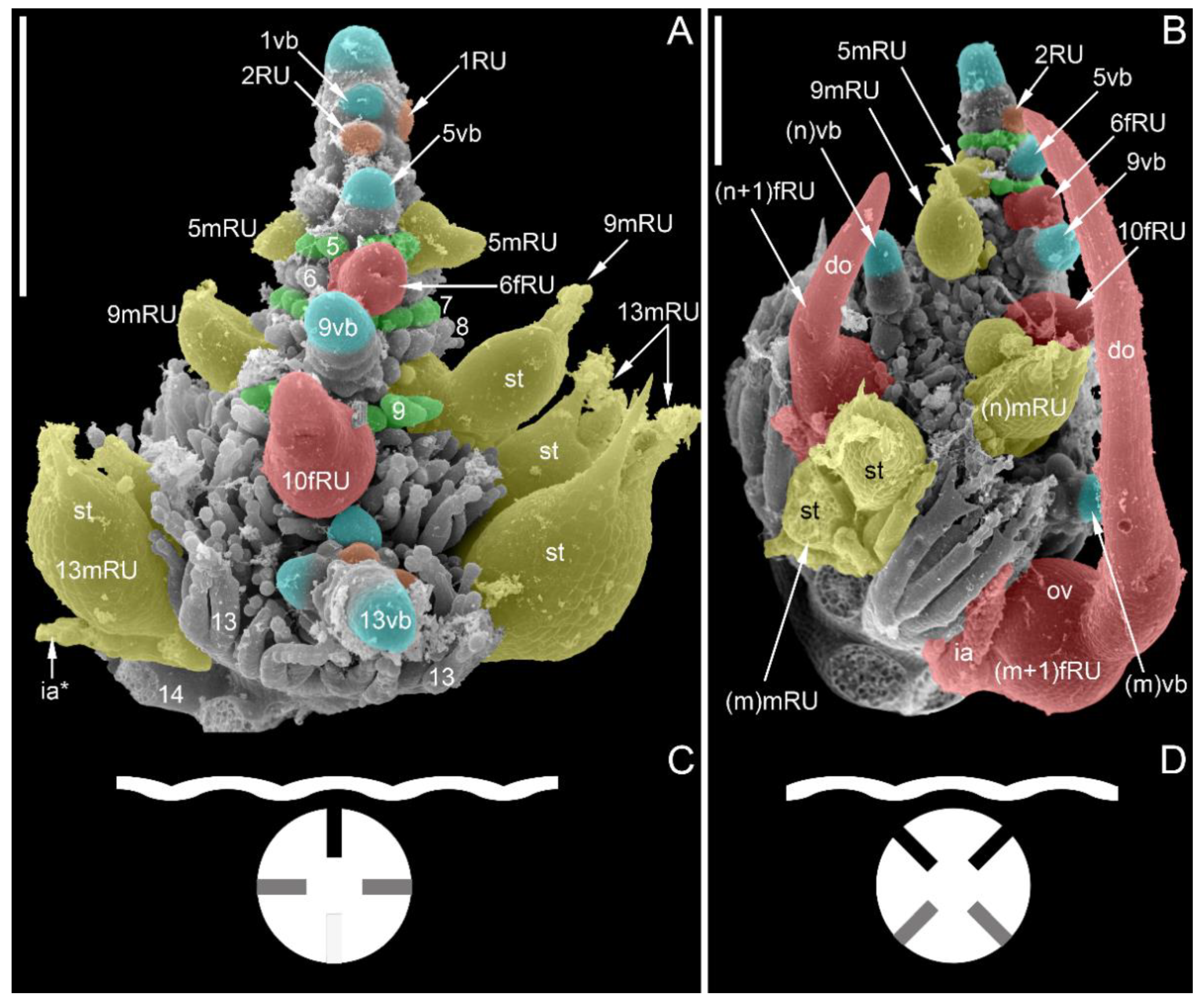
Raynal-Roques (1981) [22] described occasional occurrence of as many as four male reproductive units per node in Ceratophyllum, with two units larger than two others (Figure 2C). Raynal-Roques [22] interpreted all four male reproductive units as accessory collateral buds in the axil of one and the same leaf in the framework of the concept of dorsiventral distichy. Iwamoto et al. [25], in the framework of the decussate hypothesis, concluded that each of two leaves has a pair of collateral accessory buds. Our hypothesis implies that each node should not possess more than two reproductive units (Figure 2F). The following arguments can be used to defend our hypothesis against the problematic observation of Raynal-Roques [22]. No photographs of the nodes with four reproductive unts are available, only line drawings. None of other studies revealed this pattern. The male units are situated in closely spaced pairs, with one unit younger than the other. We speculate that the younger units may represent secondary branches of the older units. Branching of female reproductive units was documented in detail by Aboy [40]. Testing this hypothesis requires a developmental analysis of material similar to what is illustrated by Raynal-Roques [22].
(2)
Our hypothesis implies that, in some instances, out of two serial buds only one is present. When only one bud is present, this can be either a vegetative branch of a reproductive unit. Therefore, in certain instances, only an accessory bud is present and the principal bud is absent. We do not consider this point too problematic, because completely the same problem occurs with the collateral bud hypothesis with respect to shoots of Ceratophyllum tanaiticum possessing only one orthostichy of lateral axes (Figure 7).

5. Conclusions

Shoot morphology of the three species of Ceratophyllum investigated here follows certain regularities and largely corresponds to the ‘full general diagram’ (Figure 2A) with more or less significant deviations. Such fundamental regularities are the arrangement of lateral axes (vegetative branches and reproductive units) in a maximum of four orthostichies, as well as a regular alternation of vegetative branches and reproductive units along an orthostichy and among adjacent orthostichies in each node. We suggest that these more constant regularities observed in the present and earlier studies are caused by an influence of certain endogenous developmental factors responsible for organ pre-patterning in shoot apical meristem and general determination of positions where one or another type of lateral organ will arise. The actual morphology of a given shoot is also dependent on environmental factors such as shoot position relative to the water surface.
Our data support the idea that the phyllotaxis of Ceratophyllum is essentially decussate with appendages of stipular origin resembling leaf blades. We conclude that a leaf axil of Ceratophyllum possesses a complex of two serial buds, the lower producing a vegetative branch and the upper developing a reproductive unit. The reproductive unit is congenitally displaced to the subsequent node, a phenomenon known as concaulescence. Either member of the serial bud complex may be absent. Serial buds and concaulescence are known in Hedyosmum (Chloranthaceae). Our new interpretation facilitates its comparisons with Ceratophyllum.
Further research will aim to generate and analyze quantitative data on patterns of organ arrangement in shoots of Ceratophyllum. Analyses of such quantitative data were successful in another group of ancient angiosperm aquatics, Nymphaeaceae [21,70]. They supported the occurrence of certain periodicity combined with stochasticity in activity of shoot apical meristem, though no obvious adaptive function of this periodicity can be traced. A hypothesis on possible developmental mechanisms of its regulation [21] requires experimental testing. Future quantitative analyses in Ceratophyllum should allow testing ideas on periodicity of activity of shoot apical meristem, such as the occurrence of four-node [23] or three-node [23] cycles. There is a hypothesis that this periodicity facilitates an equilibrated floating of the whole branched shoot system in Ceratophyllum [23], but it requires an experimental proof. Generating quantitative data in Nymphaeaceae, especially in Nuphar, is much easier than in Ceratophyllum. Rhizomes of Nuphar retain well-recognizable scars of all lateral organs during several seasons, so that precise diagrams with more than 100 nodes can be generated [21]. Apparently, quantitative data on Ceratophyllum should be acquired through accumulation of numerous short series similar to those presented here in Figure 5.

Author Contributions

Conceptualization, methodology, and investigation, D.D.S., E.S.E. and M.V.R.; Writing—original draft preparation, D.D.S.; Writing—review and editing, E.S.E. and M.V.R. All authors have read and agreed to the published version of the manuscript.

Funding

This research was funded by the Russian Foundation for Basic Research, grant No. 18-04-00797 (shoot structure and development) and by a budgetary subsidy to the Lomonosov Moscow State University No. 121032500084-6 (the new interpretation of shoot branching).

Institutional Review Board Statement

Not applicable.

Informed Consent Statement

Not applicable.

Data Availability Statement

The original contributions presented in the study are included in the article, further inquiries can be directed to the corresponding author.

Acknowledgments

We are grateful to E.V. Pechenyuk for fixed samples of C. submersum and C. tanaiticum and to all reviewers for evaluation of our manuscript. The use of SEM was greatly facilitated by the assistance of the staff of the Laboratory of Electron microscopy at the Faculty of Biology, Lomonosov Moscow State University.

Conflicts of Interest

The authors declare no conflict of interest.

References

  1. Gatsuk, L.E. Gemmaxillary plants and a system of subordinated units of their shoot body. Bull. Mosc. Soc. Nat. Biol. Ser. 1974, 79, 100–113. [Google Scholar]
  2. Doyle, J.A. Phylogenetic analyses and morphological innovations in land plants. Annu. Plant Rev. 2013, 45, 5. [Google Scholar]
  3. Bierhorst, D.W. Morphology of Vascular Plants; Macmillan: New York, NY, USA, 1971. [Google Scholar]
  4. Bell, A.D.; Bryan, A. Plant Form: An Illustrated Guide to Flowering Plant Morphology; Timber Press: London, UK; Portland, OR, USA, 2008. [Google Scholar]
  5. Weberling, F. Morphology of Flowers and Inflorescences; Cambridge University Press: Cambridge, UK, 1989. [Google Scholar]
  6. Baum, D.A.; Day, C.D. Cryptic bracts exposed: Insights into the regulation of leaf expansion. Dev. Cell 2004, 6, 318–319. [Google Scholar] [CrossRef] [Green Version]
  7. Penin, A.A. Bract reduction in Cruciferae: Possible genetic mechanisms and evolution. Wulfenia 2008, 15, 63–73. [Google Scholar]
  8. Endress, P.K.; Doyle, J.A. Reconstructing the ancestral angiosperm flower and its initial specializations. Am. J. Bot. 2009, 96, 22–66. [Google Scholar] [CrossRef]
  9. Remizowa, M.V.; Sokoloff, D.D.; Rudall, P.J. Patterns of bract reduction in racemose inflorescences of early-divergent monocots. In Early Events in Monocot Evolution; Wilkin, P., Mayo, S.J., Eds.; Cambridge University Press: Cambridge, UK, 2013; pp. 185–207. ISBN 978-1-139-00295-0. [Google Scholar]
  10. Stevenson, D.W. Observations on vegetative branching in cycads. Int. J. Plant Sci. 2020, 181, 564–580. [Google Scholar] [CrossRef]
  11. Raciborski, M. Die Morphologie der Cabombeen und Nymphaeaceen. Flora 1894, 78, 244–279. [Google Scholar]
  12. Raciborski, M. Beiträge zur Kentniss der Cabombeen und Nymphaeaceen. Flora 1894, 79, 92–108. [Google Scholar]
  13. Cutter, E. Studies of morphogenesis in the Nymphaeaceae. I. Introduction: Some aspects of the morphology of Nuphar lutea (L.) Sm. and Nymphaea alba L. Phytomorphology 1957, 7, 45–56. [Google Scholar]
  14. Cutter, E.G. The inception and distribution of flowers in the Nymphaeaceae. Proc. Linn. Soc. Lond. 1961, 172, 93–100. [Google Scholar] [CrossRef]
  15. Richardson, F.C. Morphological Studies of the Nymphaeaceae. IV. Structure and Development of the Flower of Brasenia schreberi Gmel; University of California Press: Berkeley, CA, USA, 1969; Volume 47, pp. 1–101. [Google Scholar]
  16. Moseley, M.F. Morphological studies of the Nymphaeaceae. VI. Development of the flower of Nuphar. Phytomorphology 1971, 21, 253–283. [Google Scholar]
  17. Schneider, E.L.; Tucker, S.C.; Williamson, P.S. Floral development in the Nymphaeales. Int. J. Plant Sci. 2003, 164, S279–S292. [Google Scholar] [CrossRef]
  18. Grob, V.; Moline, P.; Pfeifer, E.; Novelo, A.R.; Rutishauser, R. Developmental morphology of branching flowers in Nymphaea prolifera. J. Plant Res. 2006, 119, 561–570. [Google Scholar] [CrossRef] [Green Version]
  19. Rudall, P.J.; Sokoloff, D.D.; Remizowa, M.V.; Conran, J.G.; Davis, J.I.; Macfarlane, T.D.; Stevenson, D.W. Morphology of Hydatellaceae, an anomalous aquatic family recently recognized as an early-divergent angiosperm lineage. Am. J. Bot. 2007, 94, 1073–1092. [Google Scholar] [CrossRef] [Green Version]
  20. Sokoloff, D.D.; Remizowa, M.V.; Briggs, B.G.; Rudall, P.J. Shoot architecture and branching pattern in perennial Hydatellaceae (Nymphaeales). Int. J. Plant Sci. 2009, 170, 869–884. [Google Scholar] [CrossRef]
  21. El, E.S.; Remizowa, M.V.; Sokoloff, D.D. Developmental flower and rhizome morphology in Nuphar (Nymphaeales): An interplay of chaos and stability. Front. Cell Dev. Biol. 2020, 8, 303. [Google Scholar] [CrossRef]
  22. Raynal-Roques, A. Contribution à L’étude Biomorphologique des Angiospermes Aquatiques Tropicales: Essai D’analyse De l’évolution. Ph.D. Thesis, Université des Sciences et Techniques du Languedoc, Montpellier, France, 1981. [Google Scholar]
  23. Rutishauser, R.; Sattler, R. Complementarity and heuristic value of contrasting models in structural botany. II. Case study on leaf whorls: Equisetum and Ceratophyllum. Bot. Jahrb. Syst. 1987, 109, 227–255. [Google Scholar]
  24. Rutishauser, R. Polymerous leaf whorls in vascular plants: Developmental morphology and fuzziness of organ identities. Int. J. Plant Sci. 1999, 160, S81–S103. [Google Scholar] [CrossRef] [Green Version]
  25. Iwamoto, A.; Izumidate, R.; Ronse De Craene, L.P. Floral anatomy and vegetative development in Ceratophyllum demersum: A morphological picture of an “unsolved” plant. Am. J. Bot. 2015, 102, 1578–1589. [Google Scholar] [CrossRef] [Green Version]
  26. Gitzendanner, M.A.; Soltis, P.S.; Wong, G.K.-S.; Ruhfel, B.R.; Soltis, D.E. Plastid phylogenomic analysis of green plants: A billion years of evolutionary history. Am. J. Bot. 2018, 105, 291–301. [Google Scholar] [CrossRef]
  27. Leebens-Mack, J.H.; Barker, M.S.; Carpenter, E.J.; Deyholos, M.K.; Gitzendanner, M.A.; Graham, S.W.; Grosse, I.; Li, Z.; Melkonian, M.; Mirarab, S.; et al. One thousand plant transcriptomes and the phylogenomics of green plants. Nature 2019, 574, 679–685. [Google Scholar] [CrossRef] [Green Version]
  28. Albert, V.A.; Renner, T. Aquatic angiosperm ambiguities answered. Nat. Plants 2020, 6, 181–183. [Google Scholar] [CrossRef] [PubMed]
  29. Mavrodiev, E.V.; Williams, D.M.; Ebach, M.C.; Mavrodieva, A.E. Fassettia, a new North American genus of family Ceratophyllaceae: Evidence based on cladistic analyses of current molecular data of Ceratophyllum. Aust. Syst. Bot. 2021, 34, 431–437. [Google Scholar] [CrossRef]
  30. Szalontai, B.; Stranczinger, S.; Mesterházy, A.; Scribailo, R.W.; Les, D.H.; Efremov, A.N.; Jacono, C.C.; Kipriyanova, L.M.; Kaushik, K.; Laktionov, A.P.; et al. Molecular phylogenetic analysis of Ceratophyllum L. taxa: A new perspective. Bot. J. Linn. Soc. 2018, 188, 161–172. [Google Scholar] [CrossRef]
  31. Guo, X.; Fang, D.; Sahu, S.K.; Yang, S.; Guang, X.; Folk, R.; Smith, S.A.; Chanderbali, A.S.; Chen, S.; Liu, M.; et al. Chloranthus genome provides insights into the early diversification of angiosperms. Nat. Commun. 2021, 12, 6930. [Google Scholar] [CrossRef]
  32. Doyle, J.A.; Endress, P.K. Phylogenetic analyses of Cretaceous fossils related to Chloranthaceae and their evolutionary implications. Bot. Rev. 2018, 84, 156–202. [Google Scholar] [CrossRef] [Green Version]
  33. Kvaček, J.; Doyle, J.A.; Endress, P.K.; Daviero-Gomez, V.; Gomez, B.; Tekleva, M. Pseudoasterophyllites cretaceus from the Cenomanian (Cretaceous) of the Czech Republic: A possible link between Chloranthaceae and Ceratophyllum. Taxon 2016, 65, 1345–1373. [Google Scholar] [CrossRef] [Green Version]
  34. Gomez, B.; Daviero-Gomez, V.; Coiffard, C.; Barral, A.; Martín-Closas, C.; Dilcher, D.L. Montsechia vidalii from the Barremian of Spain, the earliest known submerged aquatic angiosperm, and its systematic relationship to Ceratophyllum. Taxon 2020, 69, 1273–1292. [Google Scholar] [CrossRef]
  35. Antonov, A.S.; Troitsky, A.V.; Samigullin, T.K.; Bobrova, V.K.; Valiejo-Roman, K.M.; Martin, W. Early events in the evolution of angiosperms deduced from Cp RDNA ITS 2–4 sequence comparisons. In Proceedings of the International Symposium on the Family Magnoliaceae; Liu, Y.-H., Fan, H.-M., Chen, Z.-Y., Wu, Q.-G., Zeng, Q.-W., Eds.; Science Press: Beijing, China, 2000; pp. 210–214. [Google Scholar]
  36. Moore, M.J.; Hassan, N.; Gitzendanner, M.A.; Bruenn, R.A.; Croley, M.; Vandeventer, A.; Horn, J.W.; Dhingra, A.; Brockington, S.F.; Latvis, M.; et al. Phylogenetic analysis of the plastid inverted repeat for 244 species: Insights into deeper-level angiosperm relationships from a long, slowly evolving sequence region. Int. J. Plant Sci. 2011, 172, 541–558. [Google Scholar] [CrossRef] [Green Version]
  37. Zeng, L.; Zhang, Q.; Sun, R.; Kong, H.; Zhang, N.; Ma, H. Resolution of deep angiosperm phylogeny using conserved nuclear genes and estimates of early divergence times. Nat. Commun. 2014, 5, 4956. [Google Scholar] [CrossRef] [Green Version]
  38. Xue, J.-Y.; Dong, S.-S.; Wang, M.-Q.; Song, T.-Q.; Zhou, G.-C.; Li, Z.; de Peer, Y.V.; Shao, Z.-Q.; Wang, W.; Chen, M.; et al. Mitochondrial genes from 18 angiosperms fill sampling gaps for phylogenomic inferences of the early diversification of flowering plants. J. Syst. Evol. 2021; Early View. [Google Scholar] [CrossRef]
  39. Strasburger, E. Ein Beitrag zur Kenntnis von Ceratophyllum submersum und phylogenetische Erörterungen. Jahrb. Wiss. Bot. 1902, 37, 477–526. [Google Scholar]
  40. Aboy, H.E. A Study of the Anatomy and Morphology of Ceratophyllum demersum. Master’s Thesis, Cornell University, Ithaca, NY, USA, 1936. [Google Scholar]
  41. Sehgal, A.; Ram, H.Y.M. Comparative developmental morphology of two populations of Ceratophyllum L. (Ceratophyllaceae) and their taxonomy. Bot. J. Linn. Soc. 1981, 82, 343–356. [Google Scholar] [CrossRef]
  42. Shamrov, I.I. Anthecological investigation of three species of the genus Ceratophyllum (Ceratophyllaceae). Bot. Zhurnal 1983, 68, 1357–1366. [Google Scholar]
  43. Les, D.H. Systematics and Evolution of Ceratophyllum L. (Ceratophyllaceae): A Monograph (Taxonomy, Flavonoids, Aquatic plants, Phytogeography, Variation). Ph.D. Thesis, The Ohio State University, Ann Arbor, MI, USA, 1986. [Google Scholar]
  44. Endress, P.K. Evolutionary aspects of the floral structure in Ceratophyllum. Plant Syst. Evol. 1994, 8, 175–183. [Google Scholar] [CrossRef]
  45. Endress, P.K. The flowers in extant basal angiosperms and inferences on ancestral flowers. Int. J. Plant Sci. 2001, 162, 1111–1140. [Google Scholar] [CrossRef]
  46. Iwamoto, A.; Shimizu, A.; Ohba, H. Floral development and phyllotactic variation in Ceratophyllum demersum (Ceratophyllaceae). Am. J. Bot. 2003, 90, 1124–1130. [Google Scholar] [CrossRef] [PubMed]
  47. Endress, P.K.; Doyle, J.A. Ancestral traits and specializations in the flowers of the basal grade of living angiosperms. Taxon 2015, 64, 1093–1116. [Google Scholar] [CrossRef]
  48. Schaeppi, H. Untersuchungen über die Blattentwicklung bei Ceratophyllum, Cabomba und Limnophila. Planta 1935, 24, 755–769. [Google Scholar] [CrossRef]
  49. Shamrov, I.I. Morphological nature of gynoecium and fruit in Ceratophyllum (Ceratophyllaceae). Bot. Zhurnal 2009, 94, 938–961. [Google Scholar]
  50. Sokoloff, D.D.; El, E.S.; Pechenyuk, E.V.; Carrive, L.; Nadot, S.; Rudall, P.J.; Remizowa, M.V. Refined interpretation of the female flower in Ceratophyllum sheds fresh light on gynoecium evolution in angiosperms. Front. Cell Dev. Biol. 2022, 10, 868352. [Google Scholar] [CrossRef]
  51. Endress, P.K. Structure and relationships of basal relictual angiosperms. Aust. Syst. Bot. 2004, 17, 343–366. [Google Scholar] [CrossRef]
  52. Takhtajan, A. Systema et Phylogenia Magnoliophytorum; Moscow & Leningrad: Nauka, Russia, 1966. (In Russian) [Google Scholar]
  53. Cronquist, A. An Integrated System of Classification of Flowering Plants; Columbia University Press: New York, NY, USA, 1981. [Google Scholar]
  54. Goebel, K.E. Über die Verzweigung dorsiventraler Sprosse. Arb. Bot. Inst. Wuerzb. 1882, 2, 353–436. [Google Scholar]
  55. Schneider, O.H. Zur Kenntnis dorsiventraler Blütenstände in der Familie der Papilionaceen. Bot. Arch. 1925, 11, 26–39. [Google Scholar]
  56. Charlton, W.A. Pendulum symmetry. In Symmetry in Plants; Barabé, D., Jean, R.V., Eds.; World Scientific: Singapore, 1998; pp. 61–87. ISBN 978-981-02-2621-3. [Google Scholar]
  57. Loiseau, J.E.; Grangeon, D. Variations phyllotaxiques chez Ceratophyllum demersum L. et Hippuris vulgaris L. Bull. Soc. Bot. Fr. 1963, 110, 76–91. [Google Scholar] [CrossRef] [Green Version]
  58. Serebryakova, T.I. Shoot Morphogenesis and the Evolution of Life Forms in Grasses; Nauka: Moscow, Russia, 1971. [Google Scholar]
  59. Endress, P.K. Disentangling confusions in inflorescence morphology: Patterns and diversity of reproductive shoot ramification in angiosperms. J. Syst. Evol. 2010, 48, 225–239. [Google Scholar] [CrossRef]
  60. Endress, P.K.; Lorence, D.H. Inflorescence structure in Laurales—Stable and flexible patterns. Int. J. Plant Sci. 2020, 181, 267–283. [Google Scholar] [CrossRef] [Green Version]
  61. Endress, P.K.; Igersheim, A. The reproductive structures of the basal angiosperm Amborella trichopoda (Amborellaceae). Int. J. Plant Sci. 2000, 161, S237–S248. [Google Scholar] [CrossRef]
  62. Skutch, A.F. Peculiarities in the structure of the stem, related to the leaf-sheath, in Hedyosmum. Ann. Bot. 1927, 41, 715–730. [Google Scholar] [CrossRef]
  63. Todzia, C.A. Four new species of Hedyosmum (Chloranthaceae) from South America. Syst. Bot. 1988, 13, 21–31. [Google Scholar] [CrossRef]
  64. Todzia, C.A. Chloranthaceae: Hedyosmum. Flora Neotrop. 1988, 48, 1–138. [Google Scholar]
  65. Todzia, C.A.; Wood, C.E. The genus Hedyosmum (Chloranthaceae) in the West Indies. J. Arnold Arbor. 1988, 69, 51–63. [Google Scholar] [CrossRef]
  66. Doria, M.G.; Pabón-Mora, N.; González, F. Reassessing inflorescence and floral morphology and development in Hedyosmum (Chloranthaceae). Int. J. Plant Sci. 2012, 173, 735–750. [Google Scholar] [CrossRef]
  67. Sender, L.M.; Doyle, J.A.; Upchurch, G.R., Jr.; Endress, P.K.; Villanueva-Amadoz, U.; Diez, J.B. Evidence on vegetative and inflorescence morphology of Chloranthaceae (Angiospermae) from the Early Cretaceous (middle–late Albian) of Spain. J. Systemat. Palaeontol. 2021, 18, 2015–2042. [Google Scholar] [CrossRef]
  68. Ascarina lucida—The University of Auckland. Available online: https://www.nzplants.auckland.ac.nz/en/about/seed-plants-flowering/chloranthaceae/ascarina-lucida.html (accessed on 14 March 2022).
  69. D’Arcy, W.G.; Liesner, R.L. Hedyosmum (Chloranthaceae) in Panama. Syst. Bot. 1981, 6, 74–86. [Google Scholar] [CrossRef]
  70. Dormer, K.J.; Cutter, E.G. On the arrangement of flowers on the rhizomes of some Nymphaeaceae. New Phytol. 1959, 58, 176–181. [Google Scholar] [CrossRef]
Figure 1. General view of shoot system of Ceratophyllum demersum. la, leafy appendage that apparently has a stipular origin; la*, leafy appendage apparently corresponding to the leaf blade; ma, main axis; mRU, male reproductive unit; vb, vegetative branch.
Symmetry 14 01288 g001
Figure 2. Generalized roll-out diagrams of organ arrangement in shoots of Ceratophyllum. The diagrams are prepared in a special way. These are schematical side views of shoots. Each shoot is ‘cut’ longitudinally and then ‘unrolled’ to place all organ positions in the same plane. (A,B) Interpretation assuming a decussate phyllotaxis [25,48]. (A) Diagram with all available positions occupied by reproductive units and vegetative branches (‘full general diagram’). These two types of lateral axes form four orthosichies. (B) Pattern that differs in the occurrence of only two orthostichies. (C,D) Interpretation assuming a dorsiventral kind of distichous phyllotaxis [22]. (C) Pattern with all vegetative branches in two adjacent orthostichies and two, rarely four, reproductive units per node. The reproductive units are interpreted as developing from collateral buds. (D) The same pattern as in (B) under the concept of dorsiventral distichy. (E) A morphological interpretation of the branching pattern in Ceratophyllum according to ideas of Iwamoto et al. [25]. (F) A morphological interpretation of the branching pattern in Ceratophyllum proposed here. Different shades of green = leafy appendages interpreted as parts of leaves forming different orthostichies. There are four orthostichies in (A,B,E,F) and two orthostichies in (C,D). White asterisk = leafy appendage corresponding to the leaf blade (or median leaflet). Orange triangles = male or female reproductive units. Blue stars = vegetative branches. Black dotted arrows = hypothetic shifts of positions of reproductive units proposed in the two contrasting morphological interpretations in (E,F).
Symmetry 14 01288 g002
Figure 3. Patterns of organ arrangement in young seed plumule of Ceratophyllum submersum (SEM). (A) Side view of seed embryo showing a root pole that does not produce any root; (B) Oblique top view showing a plumule; (C) Top view of the embryo. cot, cotyledon; la, leafy appendage that apparently has a stipular origin; la*, leafy appendage apparently corresponding to the leaf blade; lf, leaf of the first node above the cotyledons; rp, root pole of the embryo. Scale bars = 100 μm (AC).
Symmetry 14 01288 g003
Figure 4. Patterns of organ arrangement at the beginning of vegetative branches in Ceratophyllum submersum (SEM). (A) Branch with organs of the two first nodes initiated. They can be interpreted as decussate leaves, each bearing a pair of stipules resembling the leaf blade; (B) Branch with organs of the three first nodes initiated. Each leaf can be interpreted as having a blade and four additional leafy appendages apparently of stipular origin. la, leafy appendage that apparently has a stipular origin; la*, leafy appendage apparently corresponding to the leaf blade; mRU, apex of developing male reproductive unit; vb, apex of vegetative branch; vb*, next order vegetative branch. Scale bars = 100 μm (A,B).
Symmetry 14 01288 g004
Figure 5. Roll-out diagrams of organ arrangement along mature shoots of Ceratophyllum submersum. Shoot nodes are numbered from younger to older ones in the same way as in subsequent figures. Arrows, instances of minor shifts in positions of particular orthostichies of lateral axes; dashed line, a virtual boundary between two parts of the shoot with patterns of organ arrangement not matching each other. (A,B) Shoots with minor shifts in organ positions along one orthostichy. (A) Shoot with an apparent change of a leading orthostichy between nodes 8 and 9. (B) Shoot with one stable leading orthostichy bearing vegetative branches and female reproductive units. (C) Shoot with a major shift in patterns of arrangement of lateral axes. The parts above and below the dashed line cannot be combined with each other under the concept of decussate phyllotaxis. (D) An example of orthodox branching pattern that is entirely congruent with the ‘full general diagram’ (Figure 2A).
Symmetry 14 01288 g005
Figure 6. Two views of a shoot with lateral axes arranged in three orthostichies in Ceratophyllum submersum (SEM). Nodes 2,3; then 6,7; then 10,11 do not possess lateral axes (vegetative branches or reproductive units). Note a gap (asterisk) between leafy appendages in Node 11, just above the (removed) vegetative branch. ia, involucral appendage of female reproductive unit; fRU, female reproductive unit; mRU, male reproductive unit; RU, young reproductive unit (gender yet unknown); st, stamen; vb, vegetative branch. Arabic figures, numbers of nodes where particular organs are attached (naked figures are leafy appendages). Scale bars = 100 μm (A,B).
Symmetry 14 01288 g006
Figure 7. Patterns of organ arrangement near shoot apex in Ceratophyllum submersum (SEM). Two images of a shoot with almost all lateral axes belong to one and the same orthostichy. An exception is a male reproductive unit located on the reverse side of the specimen in the Node 9 (not shown). Nodes 3,4; then 7,8; then 12 do not possess lateral axes. A vegetative branch in the Node 11 is strongly retarded in development and slightly displaced from the orthostichy, possibly by mechanical pressure of the female reproductive unit in the Node 10. fRU, female reproductive unit; or, orifice of gynoecium; ov, ovary; RU, young reproductive unit (gender yet unknown); vb, vegetative branch. Arabic figures, numbers of nodes where particular organs are attached (naked figures are leafy appendages). Scale bars = 100 μm (A,B).
Symmetry 14 01288 g007
Figure 8. Organ arrangement near shoot apex and structure of male reproductive units in Ceratophyllum submersum (SEM). (A,B) Two views of distal portion of a shoot; (C) Male reproductive unit with outermost organs removed. (D,E) Dissected male reproductive unit; (D) General view; (E) Close up of its central part showing stamens at various stages of early development. do, distal outgrowth of gynoecium; ia, involucral appendage of female reproductive unit; ia*, involucral appendage of male reproductive unit; fRU, female reproductive unit; mRU, male reproductive unit; or, orifice of gynoecium; ov, ovary; RU, young reproductive unit (gender yet unknown); st, stamen; vb, vegetative branch; Arabic figures, numbers of nodes where particular organs are attached (naked figures are leafy appendages). Scale bars = 100 μm (AE).
Symmetry 14 01288 g008
Figure 9. Leaf morphology of Ceratophyllum tanaiticum (SEM). (A) Two leafy appendages (foliage ‘leaves’) at midstage of development, each twice bifurcating. Apical glands had not released mucilage at that time; (B) Segment of leafy appendage after release of mucilage from the apical gland; (C) Vegetative branch situated below male reproductive unit and suppressed on early developmental stage. Two lateral prophylls are visible; (D) Vegetative branch with two prophylls, each developing an apical gland; (E,F) Two views of main shoot node bearing seven fully developed leafy appendages and female reproductive unit. Only proximal parts of leafy appendages (below the first bifurcation) are visible. All appendages of the node are basally united. do, distal outgrowth of gynoecium; ia, involucral appendage (of male reproductive unit in C, of female reproductive unit in E,F); la, leafy appendage; mg, mucilaginous gland; ov, ovary; st, stamen; asterisk, vegetative branch apex. Scale bars = 100 μm (AF).
Symmetry 14 01288 g009
Figure 10. Two views of a young shoot with lateral axes arranged along three orthostichies in Ceratophyllum tanaiticum (SEM). All vegetative branches are in the central orthostichy. All reproductive units of the central orthostichy are female. Digital coloring: yellow, male reproductive unit (stamen(s) plus involucral appendages); red, female reproductive unit (pistil plus involucral appendages); orange, very young reproductive unit with gender yet unknown; cyan, vegetative shoot apex; green, vegetative leaves. Vegetative leaves are colored in every second node. Leaves of proximal nodes are not colored because they are too crowded. ia, involucral appendage of female reproductive unit; ia*, involucral appendage of male reproductive unit; fRU, female reproductive unit; mRU, male reproductive unit; RU, young reproductive unit (gender yet unknown); st, stamen; vb, vegetative branch; Arabic figures, numbers of nodes where particular organs are attached numbered starting from the shoot apex (naked figures are leafy appendages). Scale bars = 300 μm (A), 100 μm (B).
Symmetry 14 01288 g010
Figure 11. Patterns of organ arrangement near a shoot apex in Ceratophyllum tanaiticum (SEM). (A) Shoot with female reproductive units and vegetative branches arranged along the same (central) orthostichy. It is called here ‘leading orthostichy’ and most likely positioned towards the water surface. The left and right orthostichies have male reproductive units only; (B) Shoot with lateral axes arranged along four orthostichies, but only two are visible in the view; the two orthostichies not visible contain male reproductive units only. Digital coloring in (A,B): yellow, male reproductive unit (stamen(s) plus involucral appendages); red, female reproductive unit (pistil plus involucral appendages); orange, very young reproductive unit with gender yet unknown; cyan, vegetative shoot apex; green, vegetative leaves. Vegetative leaves are colored in every second node. Leaves of proximal nodes are not colored because they are too crowded. do, distal outgrowth of gynoecium; ia, involucral appendage of female reproductive unit; ia*, involucral appendage of male reproductive unit; fRU, female reproductive unit; mRU, male reproductive unit; ov, ovary; RU, young reproductive unit (gender yet unknown); st, stamen; vb, vegetative branch; Arabic figures, numbers of nodes where particular organs are attached (naked figures are leafy appendages). The nodes are numbered starting from the shoot apex. Nodes are difficult to count in the proximal part of B and arbitrary node numbers n and m are thus introduced; the number n is probably 12. Scale bars = 300 μm (A,B). (C,D) Possible link between the shoot orientation relative to water surface and the number of leading orthostichies. (C) Shoot with single leading orthostichy as in (A); (D) Shoot with two leading orthostichies as in (B). Wavy line, water surface; white circle, diagrammatic cross-section of stem; black lines, positions of leading orthostichies; grey lines, positions of other orthostichies of lateral axes.
Symmetry 14 01288 g011
Figure 12. A shoot of Ceratophyllum tanaiticum with a change of leading orthostichy (SEM). The shoot is shown in two views (A,B). Most lateral axes belong to two orthostichies. These are labeled blue and magenta. In younger part of the shoot, the blue orthostichy is a leading one and bears vegetative branches as well as female reproductive units. In older part of the shoot, the magenta orthostichy is a leading one and bears vegetative branches as well as female reproductive units. Male reproductive units belong to non-leading parts of the blue and magenta orthostichies. There are two more male reproductive units (one shown in (A) and the other in (B)) that belong to their own orthostichies. Arabic figures, numbers of main axis nodes to which particular organs belong; naked figures are leafy appendages. Precise numbering is problematic after the Node 6. fRU, female reproductive unit; mRU, male reproductive unit; RU, reproductive unit (gender yet unknown); vb, vegetative branch. Scale bars = 100 μm (A,B).
Symmetry 14 01288 g012
Figure 13. Ceratophyllum tanaiticum (SEM), young organs below the shoot apex. (A) Axillary vegetative branch is initiated in Node 3; non-axillary reproductive unit is initiated in the same orthostichy in Node 4; (B) Shoot with reproductive units present in every node. Reproductive units in nodes 1–3 are yet undifferentiated, their gender is unknown. Reproductive units in nodes 4 and 5 are female, each having a female flower surrounded by involucral appendages. Development of reproductive units is not acropetal. The reproductive unit in the Node 3 is smaller than those in nodes 1 and 2. Out of two reproductive units of the Node 4, one is smaller and another is larger than the reproductive unit in the Node 5. ff, female flower; ia, involucral appendage; RU, young reproductive unit (gender yet unknown); vb, vegetative branch; Arabic figures, numbers of nodes where particular organs are attached; asterisk, leafy appendage subtending a vegetative branch. Scale bars = 30 μm.
Symmetry 14 01288 g013
Figure 14. Development of male reproductive units in Ceratophyllum tanaiticum (SEM). (A) Detail of shoot with very young male reproductive unit and vegetative branch at the same node. The male reproductive unit can be recognized by its monosymmetric shape due to first evidence of appearance of the abaxial stamen; (B) Male reproductive unit with abaxial stamen initiated. A massive reproductive unit apex is left after the stamen initiation; (C) The second, adaxial stamen is initiated and no residual reproductive unit apex is left; involucral appendages are initiated; (D) Male reproductive unit that will apparently remain one-staminate, because the stamen is older than st1 in C, but the second stamen is not initiated; (E) Top view of two-staminate RU; (F) Top view of three-staminate RU. fRU, female reproductive unit; ia, involucral appendage; mg, mucilaginous gland; mRU, male reproductive unit; mRUa, male reproductive unit apex; st, stamens (numbered in sequence of initiation); vb, vegetative branch. Arabic figures in (A), numbers of nodes where particular organs are attached; naked figures are leafy appendages. Scale bars = 30 μm (AE), 100 μm (F).
Symmetry 14 01288 g014
Figure 15. Mature male reproductive units of Ceratophyllum tanaiticum (SEM). (A) Side view of one-staminate reproductive unit; (B) Top view of one-staminate reproductive unit with the stamen removed; no residual apex of the reproductive unit can be seen at this stage; (C) Side view of three-staminate RU. an, anther; ia, involucral appendage; mg, mucilaginous gland. Scale bars = 100 μm (AC).
Symmetry 14 01288 g015
Figure 16. Patterns of organ arrangement and development in Ceratophyllum demersum (SEM). Shoots developing only vegetative branches. (A) Distal part of a shoot. Vegetative branch apex in the Node 2 is larger than that in the Node 3. (B) Young vegetative branch. The leafy appendage in its radius (3*) is much smaller than other leafy appendages of this node (3). (C) Distal part of a shoot. Vegetative branch apex in the Node 2 is larger than that in the Node 3. The branch in the Node 2 is closely associated with a small leafy appendage in its radius and the two structures likely developed from a common primordium. The branch in Node 5 is larger than that in the Node 6. vb, vegetative branch; Arabic figures, numbers of nodes where particular organs are attached; naked figures are leafy appendages apparently corresponding to stipules; figures with asterisks are leafy appendages likely corresponding to leaf blades. Scale bars = 30 μm (AC).
Symmetry 14 01288 g016
Figure 17. Structure and position of young reproductive units and vegetative branches in Ceratophyllum demersum (SEM). (A) Shoot with three vegetative branches and two reproductive units initiated. Their arrangement fits the diagram in Figure 2B; (B) Detail of another shoot most likely showing a later developmental stage of structures similar to those in the right-hand orthostichy of (A). Only gynoecium is visible in the female reproductive unit in (B), the young involucral appendages of the unit are hidden by the gynoecium and surrounding structures. la, la*, leafy appendages of the vegetative branch; la, appendages of stipular origin; la*, appendages corresponding to the leaf blade; fRU, female reproductive unit; RU, young reproductive unit (gender yet unknown); vb, vegetative branch. Arabic figures, numbers of nodes where particular organs are attached; naked figures are leafy appendages of stipular origin; figures with asterisks are those likely corresponding to leaf blades. Scale bars = 30 μm (A,B).
Symmetry 14 01288 g017
Publisher’s Note: MDPI stays neutral with regard to jurisdictional claims in published maps and institutional affiliations.

