Review

Red Sea Atlas of Coral-Associated Bacteria Highlights Common Microbiome Members and Their Distribution across Environmental Gradients—A Systematic Review

by
Nathalia Delgadillo-Ordoñez
1,†,
Inês Raimundo
1,†,
Adam R. Barno
1,
Eslam O. Osman
1,
Helena Villela
1,
Morgan Bennett-Smith
1,
Christian R. Voolstra
1,2,
Francesca Benzoni
1 and
Raquel S. Peixoto
1,*
1
Marine Microbiomes Laboratory, Red Sea Research Center (RSRC), Biological and Environmental Sciences and Engineering Division (BESE), King Abdullah University of Science and Technology (KAUST), Thuwal 23955, Saudi Arabia
2
Department of Biology, University of Konstanz, 78457 Konstanz, Germany
*
Author to whom correspondence should be addressed.
These authors contributed equally to this work.
Microorganisms 2022, 10(12), 2340; https://doi.org/10.3390/microorganisms10122340
Submission received: 28 August 2022 / Revised: 12 October 2022 / Accepted: 18 November 2022 / Published: 26 November 2022
(This article belongs to the Special Issue Microbiomes of Aquatic Organisms)

Abstract

:
The Red Sea is a suitable model for studying coral reefs under climate change due to its strong environmental gradient that provides a window into future global warming scenarios. For instance, corals in the southern Red Sea thrive at temperatures predicted to occur at the end of the century in other biogeographic regions. Corals in the Red Sea thrive under contrasting thermal and environmental regimes along their latitudinal gradient. Because microbial communities associated with corals contribute to host physiology, we conducted a systematic review of the known diversity of Red Sea coral-associated bacteria, considering geographic location and host species. Our assessment comprises 54 studies of 67 coral host species employing cultivation-dependent and cultivation-independent techniques. Most studies have been conducted in the central and northern Red Sea, while the southern and western regions remain largely unexplored. Our data also show that, despite the high diversity of corals in the Red Sea, the most studied corals were Pocillopora verrucosa, Dipsastraea spp., Pleuractis granulosa, and Stylophora pistillata. Microbial diversity was dominated by bacteria from the class Gammaproteobacteria, while the most frequently occurring bacterial families included Rhodobacteraceae and Vibrionaceae. We also identified bacterial families exclusively associated with each of the studied coral orders: Scleractinia (n = 125), Alcyonacea (n = 7), and Capitata (n = 2). This review encompasses 20 years of research in the Red Sea, providing a baseline compendium for coral-associated bacterial diversity.

1. Introduction

Corals are cnidarian hosts that live in association with several groups of microorganisms, including viruses, bacteria, archaea, dinoflagellate algae, and fungi, which together comprise the so-called holobiont [1,2]. The endosymbiotic relationship between the coral host and photosynthetic dinoflagellates (family Symbiodiniaceae) provides up to 90% of the host nutritional demands, and it is critical to sustaining reef ecosystem functioning [3,4]. In addition to Symbiodiniaceae, other microorganisms, notably prokaryotes (referred to as the microbiome hereafter), associate with corals and play an important role in coral physiology and health [5,6,7,8,9,10]. The coral microbiome is involved, among other chemical and biological processes, in carbon, nitrogen, and sulfur cycling, defense against pathogens, environmental adaptability, and degradation of toxic compounds [9,10,11,12,13,14,15,16,17]. However, the elaborated interactions between the host and its microbiome are complex, and it is yet to be fully understood how microbial communities can improve coral holobiont functions to tolerate environmental stressors, particularly those derived from ocean warming [9,13].
Coral-associated microbiomes are dynamic communities that show flexibility (e.g., restructuring of the microbial community) to changing environmental conditions as a potential adaptive mechanism for holobiont health [18,19]. Overall, these microbial communities present (i) host specificity, where each coral species has, to some extent, distinct associated microbial groups that compose species-specific microbiomes [20,21]; (ii) variable microbial groups that respond to environmental conditions [20]; and (iii) distinct communities between coral compartments, i.e., mucus, tissue, and skeleton [22,23]. As such, coral hosts are likely to select the most advantageous microbial partners that contribute to adaptation to environmental perturbations and, consequently, their survival [24,25]. Therefore, understanding the composition, diversity, and dynamics of microbiomes associated with corals across environmental settings is key to understanding the potential role of the microbiome in improving coral acclimation to environmental stressors, including those derived from ongoing ocean warming.
The Red Sea is a biodiversity hotspot that harbors thousands of marine species and is populated by extensive and healthy coral reefs, with a high rate of endemism [26,27]. Coral holobionts in the Red Sea have adapted to an overall unique marine environment characterized by a combination of conditions deemed extreme both in shallow and deep water compared to other regions. In the shallow photic zone, Red Sea corals experience salinity of 41 PSU and summer temperatures range from 25–32 °C along the latitudinal gradient [28,29]. In the aphotic zone, the water temperature never drops below 21 °C and salinity remains around 40.5 PSU. Similarly, the chlorophyll concentration is higher in the southern Red Sea due to the exchange with nutrient-rich seawater from the Gulf of Aden, and consequently, seawater visibility in the south is lower than in the central and northern Red Sea regions [30]. Despite the contrasting environmental conditions, genetic analysis of coral species have shown high gene flow and genetic similarity between coral populations along the latitudinal gradient of the Red Sea, suggesting that coral hosts have low genetic variations along the Red Sea latitudinal gradient [31,32]. Conversely, patterns of continuous latitudinal zonation have been highlighted in selected scleractinian coral hosts for the associated Symbiodiniaceae communities [33,34]. All of the above may highlight the potential of microbiome restructuring as an adaptive strategy to influence species distribution along the Red Sea to likely facilitate survival of the same coral species under adverse environmental regimes. Osman and colleagues (2020) found plasticity of microbiome communities across latitudinal gradients of the northern Red Sea, but high specificity of endosymbiotic algae [21]. As such, the Red Sea can be seen as a natural laboratory to assess the response of coral microbiomes to different environmental conditions and how microbiome flexibility could increase the coral holobiont tolerance to environmental shifts.
Despite the increasing number of microbiome studies in recent years, e.g., [10,35], the number of publications on the Red Sea remains relatively small in comparison to other regions, such as the Great Barrier Reef, and has focused only on a few coral species and/or has been restricted to certain regions [27]. Few studies have assessed the coral microbiome composition and its dynamics along its latitudinal gradient in the Red Sea, e.g., [21,36,37]. Here, we performed a systematic review of the current knowledge of the Red Sea coral associated bacteria using publicly accessible data. The objectives were: (1), to assess and summarize the existing Red Sea coral bacteria information published over the past two decades and (2), to detect current knowledge gaps to inform the direction of future fundamental and applied research. Our analysis of the published data allowed us to assess the bacterial composition of several Red Sea coral taxa. Moreover, we identify bacterial taxa associated with healthy corals and their distribution patterns along the latitudinal gradient of the Red Sea. Thus, we provide the first comprehensive atlas of coral-associated bacteria in the Red Sea, serving as a baseline for future fundamental and applied research including coral restoration projects.

2. Materials and Methods

2.1. Red Sea Studies Survey

A systematic survey for published Red Sea coral-associated bacteria was conducted according to the PRISMA guidelines [38], using the Web of Science and PubMed public databases from July 2002 until February 2021 (https://www.webofscience.com/wos/woscc/basic-search (accessed on 1 February 2021); https://pubmed.ncbi.nlm.nih.gov/ (accessed on 1 February 2021), respectively). The available literature published over the past two decades along the Red Sea was compiled using five keywords for the web search: “Red Sea”, “coral”, “bacteria”, “microbiome” and “microbes”, in their different combinations, using the restrictor “AND” to limit the search. The search produced 151 results, and publications that were not from the Red Sea were excluded. The retrieved papers were downloaded and each paper was assessed to determine whether it was relevant to the coral associated bacteria or not, excluding the publications that studied free-living marine bacteria, marine sediments, aquaculture, and marine environments different from coral reefs. Thereafter, a total of 54 studies were selected, and metadata for each paper were extracted, including the year of publication, the studied coral species, geographic location (coordinates), sampling depth, sequencing technology (i.e., culture-based vs. sequencing), and associated GenBank accession numbers. The publications and studied coral species were counted to quantify the number of coral microbiome studies along the latitudinal gradient of the Red Sea. The geographic coordinates of the sampling locations were retrieved from each study. However, for the studies that did not provide this information (n = 9), the coordinates of the sampling sites were estimated using Google Earth based on the available information in each reference. For this, coordinates were estimated by reading the location description section in the retrieved paper to locate the sampling site on Google Earth. Then, a middle point was taken as a representative coordinate for the study. Coordinates were transformed into a decimal system and plotted over the Red Sea map using ArcGIS Pro software (V 2.8.0). The Fishnet tool in ArcGIS pro (V 2.8.0) was used to quantify the number of studied sites per 100 km2 grid in the different regions of the Red Sea.

2.2. Coral-Associated Bacteria in the Red Sea

We used the data from the retrieved publications to obtain a list of bacteria associated with different coral species along the Red Sea. We focused on bacterial communities associated with healthy corals (n = 36 publications) because our objective was an assessment of the coral associated bacterial communities and their variability under natural conditions in the study area. Thus, samples detailing diseased, bleached, temperature- or nutrient-treated coral and seawater samples were removed from the analysis (n = 18 publications, see S1). Samples for which it was not possible to retrieve the data were similarly excluded. Furthermore, we only considered studies involving culture-independent techniques (n = 24 publications) for the bacterial atlas. Bacterial diversity retrieved from culture-dependent techniques was considered separately (n = 12 publications). The taxonomic profile for each sample was extracted from NCBI using the samples’ respective Sequence Read Archive (SRA) and/or accession numbers provided. Bacterial taxonomy was extracted from each unique SRA linked to 16S rRNA BioSamples using the Taxonomy Analysis tool in NCBI. The compiled taxonomic profiles for biological replicates per coral taxa within each study were aggregated and duplicate bacterial taxa were removed. Therefore, for each study, each bacterial taxon was represented only once, as presence/absence, for the studied corals. Here, we assessed the previously reported diversity of bacteria associated with corals, and did not compare and/or reanalyze the data. Therefore, in this atlas we included publications regardless of their different sequencing methods (Sanger vs. Next Generation Sequencing (NGS)), 16S rRNA gene regions (e.g., V2 vs. V3), platforms (e.g., HiSeq vs. MiSeq), pipelines (e.g., QIIME vs. mothur), and taxonomy resolution (OTUs = Operational Taxonomic Unit vs. ASVs = Amplicon Sequence Variants). The lowest taxonomic level that provided the most reliable information was the family level; hence, all the results presented were pooled to this taxonomic unit level of resolution. In addition, SRA annotations were missing in two publications [21,39]. In these cases, we used the taxonomic information provided within the publication.
The occurrence of each bacterial family was counted for all studied coral species along the Red Sea, excluding duplicates and biological replicates to avoid erroneous estimations in the data. Similarly, the reported diversity of coral bacterial communities between different Red Sea regions was assessed. For this, the Red Sea was subdivided into three regions known to have a different environmental regime [28,40,41], namely the northern, central, and southern Red Sea (lat. 29, 24, and 20, respectively). The number of reported bacterial families within each region was counted according to the available retrieved data from each publication to investigate the similarity and disparity between and among regions. In addition, we identified common bacterial families across coral species to highlight their possible roles in the holobiont in the context of future management directions and restoration applications.
The classification of the studied corals was updated to reflect the most recent taxonomic framework based on formal published revisions as per the World Register of Marine Species [42] and references therein. When outdated, the coral taxa names used in the original examined papers were modified. For example, for literature reports of Favia favus (Forskål, 1775), Fungia granulosa (Klunzinger, 1879), and Fungia scutaria (Lamarck, 1801), we adopted the most recently revised genus assignation, namely Dipsastraea favus, Pleuractis granulosa, and Lobactis scutaria, respectively. For examined samples from coral hosts lacking a deposited DNA and/or skeleton voucher, allowing re-identification of a potentially doubtful species-level assignation [43], the data were included in the analyses and cf was added to the species name to reflect such uncertainty. An abbreviation of confer in Latin, literally “compare with” (cf), is used in the taxonomic literature before a taxa name to indicate a level of uncertainty for its identification [44]. In our case, specifically, material identified as Pocillopora damicornis (Linnaeus, 1758) in the examined studies is here referred to as P. cf damicornis due to a lack of genetic identification of the examined material in the original paper. Pocillopora is a notoriously complex and diversified coral genus that poses particular challenges to in situ identification due to its colony plasticity and limited informative microstructural characteristics [45]. Lately, reliable species-level identification for this genus has, therefore, mostly been performed genetically. While Pocillopora verrucosa has been recorded as present in the Red Sea, e.g., [31], the presence of P. damicornis has not been confirmed thus far based on the published literature [46]; (Schmidt-Roach pers. comm.). Similarly, the fungiid Ctenactis echinata (Pallas, 1766) is common and locally abundant in the Red Sea [30,47,48]; however, the presence of its congener Ctenactis crassa (Dana, 1846) in the region, although reported in one of the references we examined here [49], has not yet been confirmed based on genetic data evidence. Therefore, while data from the study were still included in our analyses, the coral host is reported as C. cf crassa to reflect the aforementioned species level record uncertainty.

3. Results

3.1. Coral-Associated Bacteria Literature Survey

The assessment of the published literature retrieved on the Web of Science and PubMed databases revealed the presence of 54 studies on coral bacterial communities along the Red Sea over the past two decades (Table 1). These studies covered 152 sampling sites and the central Red Sea had the greatest number of sites (n = 85), followed by the north (n = 56), then south (n = 11) (Figure 1a). During 2000–2011, the total number of studies was 16 and this increased to 54 in the past decade (Figure 1b).
After retrieving the studies from the databases, and prior to excluding those not matching our criteria for the Red Sea bacterial atlas (see Figure 1d), the number of studied coral taxa was 67 (44 corals sensu lato classified to species and 23 to genus level). Among corals, broad term grouping representatives of multiple classes in the phylum Cnidaria, hard corals (n = 47, 70%), including scleractinians (Anthozoa, Hexacorallia, Scleractinia) and hydrocorals (Hydrozoa, Anthoathecata, Capitata), were the most represented, followed by soft corals and gorgonians (Anthozoa, Octocorallia, Alcyonacea) (n = 20, 30%). Out of 54 references, 76% (n = 41 papers) investigated the microbiome communities associated with healthy corals, whereas 17% were conducted on corals showing signs of disease (n = 9). Four studies focused on the microbial communities associated with seawater in coral reefs and/or microbial biofilms in artificial structures (Autonomous Reef Monitoring Structures “ARMS” or Terracotta Tiles) rather than on specific coral species (representing 7% of the total studies) (Figure 1c). Furthermore, culture-independent techniques were the most commonly used methods to identify the coral associated bacteria (n = 32 papers, 57%) compared to culture-dependent techniques, which were relatively less common (n = 20, 39%), while 3.7% of the studies (n = 2) used both culture-dependent and -independent approaches (Figure 1c). In addition, most of the studies focused on the photic zone (n = 52 papers, 96.3%) up to 50 m deep, whereas only 2 studies (4%) targeted deep-sea corals (aphotic zone) between 300 and 1000 m (Figure 1c). The predominant corals studied were Pocillopora verrucosa (Ellis and Solander, 1786) (n = 8 publications, 15%), followed by Favia sp. (= Dipsastraea sp.), Pleuractis granulosa (n = 7, 13%), Stylophora pistillata (Esper, 1792) (n = 6, 11%), Acropora hemprichii (Ehrenberg, 1834) (n = 4, 7%), Dipsastraea favus (Forskål, 1775), and Platygyra sp. (n = 4, 7%, each). Additional information from the retrieved studies is available in Supplementary Material S1.

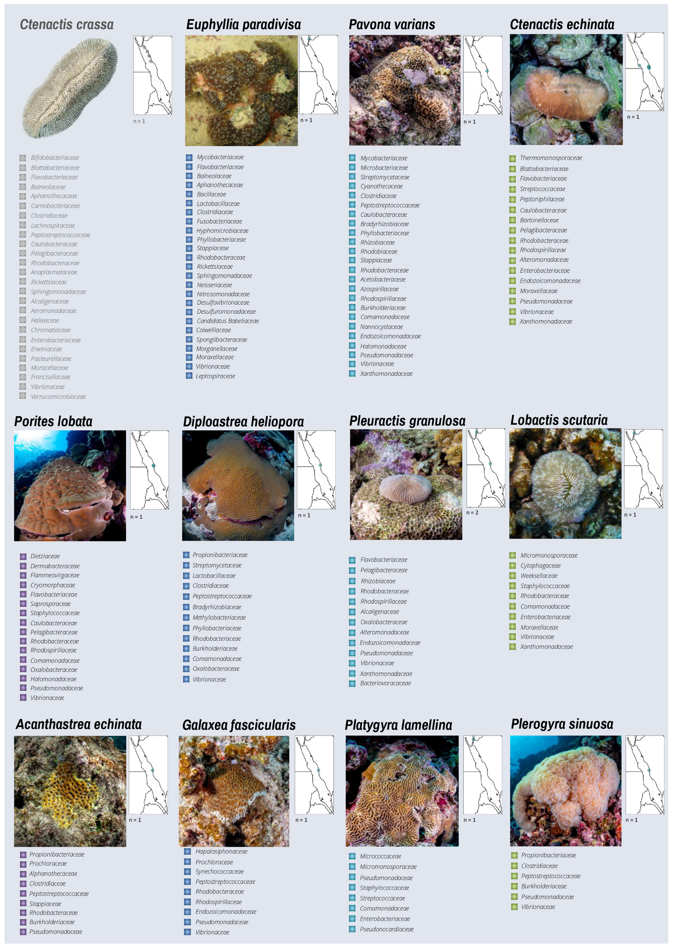
3.2. Bacterial Diversity of Red Sea Corals from Cultivation-Independent Techniques

Out of 54 retrieved studies, the 24 that investigated coral-associated bacteria with healthy corals using culture-independent techniques were included in the diversity analysis/atlas (Figure 1d). Overall, we found 43 phyla, 67 classes, 150 orders, and 324 bacterial families associated with 35 taxa of healthy corals (Supplementary Material S2). Proteobacteria was the phylum with the highest occurrence across all coral taxa (i.e., 49.8%), followed by Firmicutes (11.9%), Actinobacteria (10.4%), Bacteroidetes (10%), Acidobacteria (3.1%), Verrucomicrobia (2%), Cyanobacteria (1.9%), and Planctomycetes (1.4%), which together comprised 90.7% of the total reported phyla across all studies (Figure 2a). Gammaproteobacteria (20.4%) and Alphaproteobacteria (17%) were the most identified classes within the phylum Proteobacteria, followed by Betaproteobacteria (5.5%), Deltaproteobacteria (4.8%), and Epsilonproteobacteria (1.2%) (Figure 2b). The occurrence of further phyla, classes, and orders is detailed in Figure 2a–c and Supplementary Material S2. The families with the highest occurrence across all coral taxa and sites were Rhodobacteraceae (1.4%—Alphaproteobacteria), Pseudomonadaceae (1.3%—Gammaproteobacteria), Vibrionaceae (1.3%—Gammaproteobacteria), Flavobacteriaceae (1.2%—Bacteroidetes), Peptostreptococcaceae (1%—Clostridia), Burkholderiaceae (1%—Betaproteobacteria), Enterobacteriaceae (0.9%—Gammaproteobacteria), Moraxellaceae (0.9%—Gammaproteobacteria), Alteromonadaceae (0.8%—Gammaproteobacteria), and Xanthomonadaceae (0.8%—Gammaproteobacteria), which together comprised 10.7% of all reported families (Figure 2d). Families representing less than 0.5% of the occurrences were groupedas “Others” and comprised 52.5% of the occurrences. Unclassified families represented 21.2% of all the reported families (Supplementary Material S2).
The presence/absence of each bacterial family within each coral host taxon was counted to assess their richness and provide a descriptive survey of their associations (Figure 3, Figure 4 and Figure 5). Seven coral taxa had a higher number of reported bacterial families (n > 130) (i.e., the hard corals Stylophora pistillata, Seriatopora hystrix, Dipsastraea favus, Porites nodifera and Pocillopora cf damicornis, and the soft corals Sarcophyton trocheliophorum and Xenia umbellata). The remaining coral species presented a moderate or low number of bacterial families identified by the available publications, ranging from 76 families in Pocillopora verrucosa to 2 families in Acropora humilis (Figure 3a). Interestingly, 52 distinct bacterial families were commonly reported in 10 or more different coral taxa (Figure 3b), while others were exclusively identified in some corals. For instance, the families Rhodobacteraceae, Vibrionaceae, Pseudomonadaceae, Peptostreptococcaceae, Flavobacteriaceae, Burkholderiaceae, and Enterobacteriaceae were reported in >60% of the corals studied, considering a range of sequencing technologies, depths, and bioinformatics analyses utilized per study, while 93 families have been described in unique coral taxa of these studies (Supplementary Material S2). A descriptive Atlas of our current knowledge on the bacterial diversity (represented here at the family level) reported for 32 coral species is shown in Figure 4, in addition to the distribution of the study sites in the publications used for the Atlas per coral species along the Red Sea.

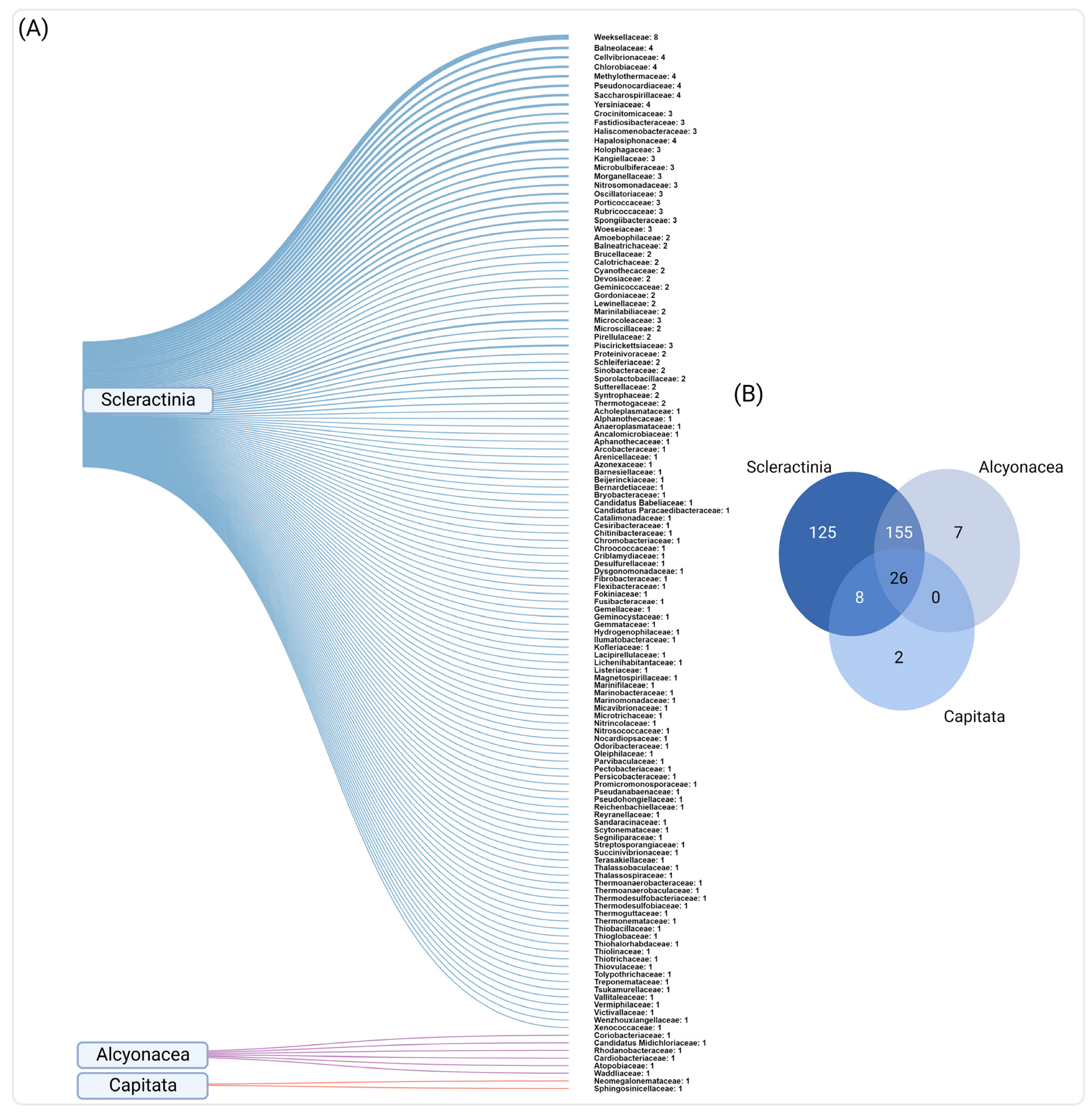
3.3. Common and Unique Bacteria in Red Sea Corals

We found several bacterial families that exist in coral host taxa regardless of their region of provenance in the Red Sea. For example, Rhodobacteraceae was identified in 30 coral taxa out of the 35 studied (85.7%), followed by Vibrionaceae (n = 29, 82.9%), Pseudomonadaceae (n = 27, 77.1%), Peptostreptococcaceae (n = 23, 65.7%), and Flavobacteriaceae (n = 23, 65.7%). Furthermore, families such as Burkholderiaceae (n = 22, 62.9%), Enterobacteriaceae (n = 21, 60%), Caulobacteraceae, Alteromonadaceae, Moraxellaceae (n = 19, 54.3% each), and Rhodospirillaceae (n = 18, 51.4%) were found in more than 50% of the coral taxa, followed by Xanthomonadaceae (n = 17, 48.6%), Comamonadaceae, Phyllobacteraceae (n = 16, 45.7% each), Staphylococcaceae, Alcaligenaceae, Sphingomonadaceae, and Streptococcaeae (n = 15, 42.8% each), which were slightly less frequent. Other families, such as Oxalobacteraceae, Propionibacteraceae, Chromatiaceae (n = 14. 40% each), Mycobacteriaceae, Streptomycetaceae, Rhizobiaceae, Lactobacillaceae, Corynebacteriaceae, Helicobacteraceae (n = 13, 37.1% each), Halomonadaceae, Clostridiaceae, Endozoicomonadaceae, Collwelliaceae, Hyphomonadaceae, and Dietziaceae (n = 12, 34.3% each) were less commonly identified but still had a comparably high occurrence across coral taxa. Other frequently found bacteria are detailed in Figure 3b.
We also explored the diversity of bacterial families associated with the three coral orders (Scleractinia, Alcyonacea, and Capitata) to investigate common and unique families reported for each coral group (Figure 5a,b). We found 26 families that were commonly associated with the three coral orders, including some of the most frequently reported families (e.g., Rhodobacteraceae, Vibrionaceae, Pseudomonadaceae, Peptostreptococcaceae, and Flavobacteraceae). Interestingly, we also found exclusive bacterial associations for each of the three groups. For instance, 125 families were exclusively associated with Scleractinia. Among them, Wekselliaceae was the most frequently reported family across corals of this group (identified eight times, corresponding to 0.3% of the total microbial identifications), followed by Yersiniaceae, Balneolaceae, Cellvibrionaceae, Chlorobiaceae, Methylothermaceae, Pseudonocardiaceae, and Saccharospirillaceae (n = 4, 0.2% each). Alcyonacea presented a lower number of exclusive families (n = 7), such as Coriobacteriaceae (n = 3, 0.1%), Elusimicrobiaceae, Rhodanobacteraceae, Cardiobacteriaceae, Atopobiaceae, Waddliaceae and Candidatus Midichloriaceae (n = 1, 0.04% each), while only two bacterial families were exclusively associated with Capitata (Neomegalonemataceae and Sphingosinicellaceae, n = 1, 0.04%) (Figure 5a,b; Supplementary Material S2).

3.4. Coral Bacteria across Red Sea Regions

The assessment of bacterial communities among the Red Sea regions, based on the 24 studies assessed, revealed a decline in the number of bacterial families along the latitudinal gradient toward the south. The northern Red Sea had the highest number of families (n = 282), followed by the central (n = 203) and southern (n = 41) regions. The families with the highest occurrence in the northern region were Rhodobacteraceae, Vibrionaceae, and Moraxellaceae (i.e., 0.7% each, of the total occurrence for this region), followed by Flavobacteriaceae (0.68%), Alteromonadaceae, Enterobacteriaceae, Pseudomonadaceae, Sphingomonadaceae, and Staphylococcaceae (0.6% each). In the central Red Sea, the most frequently identified families were Rhodobacteraceae, Pseudomonadaceae (2.8% each), Vibrionaceae (2.7%), Flavobacteriaceae (2.4%), Peptostreptococcaceae (2.2%), and Burkholderiaceae (2.1%). Finally, the families with the highest occurrence in the southern region were Endozoicomonadaceae and Rhodobacteraceae (5.5% each), Alcaligenaceae, Alteromonadaceae, Anaplasmataceae, Clostridiaceae, and Pseudomonadaceae (3.7% each) (Supplementary Material S2).
Thirty-six families were common across these regions (e.g., Rhodobacteraceae, Vibrionaceae, Flavobacteriaceae, Pseudomonadaceae, and Endozoicomonadaceae, among others). Some of these families were also the most frequently identified across coral taxa (Rhodobacteraceae, Vibrionaceae, Pseudomonadaceae, and Flavobacteriaceae), as mentioned previously. Additionally, 115 families were exclusively reported in the northern region only, 40 families in the central region, and only 3 families were found exclusively in the southern Red Sea (Thiotrichaceae, Acholeplasmataceae, Nocardiopsaceae) (Figure 3c; Supplementary Material S2).

3.5. Cultured Bacterial Diversity of Red Sea Corals

We examined the bacterial diversity from 12 studies that used culture-dependent techniques to isolate bacteria from healthy corals (Supplementary Material S3). From a total of 21 coral taxa, including hard (n = 10 coral taxa, 47.6%) and soft corals (n = 11, 52.4%), the coral with the highest number of reported bacterial families was Lobactis scutaria (n = 12), followed by Sarcophytum glaucum (n = 9), Acropora cytherea (n = 8), Pleuractis granulosa (n = 6), Stylophora sp. (n = 5), Xenia sp. (n = 4), Favia sp., Acropora hemprichii, Acropora humilis (n = 4 each), Platygyra sp. (n = 3), Porites sp., Pocillopora sp. (n = 3 each), Litophyton arboreum, Rhytisma fulvum, Lobularia sp. (n = 2 each), and Dendronephthya sinaiensis, Heteroxenia fuscescens, Dendronephthya hemprichi, Rhytisma fulvum fulvum, Sinularia sp., and Millepora dichotoma (n = 1 each) (Figure 6a).
The reported bacterial diversity was considerably lower than in the studies that used culture-independent techniques. For instance, only 4 phyla, 5 classes, 22 orders, and 33 families were reported. The isolated bacterial diversity was composed of 47 isolates that belonged to the phylum Proteobacteria (approximately 61%), followed by Actinobacteria (24.6%) and Firmicutes (12.9%), while only one belonged to Bacteroidetes (1.3%). The classes were dominated by Gammaproteobacteria (46.7%), followed by Actinomycetia (24.6%), Alphaproteobacteria (14.3%), Bacilli (12.9%), and Cytophagia (1.3%). Orders were dominated by Alteromonadales (15.6%), Bacillales (12.9%) and Vibrionales (10.4%), followed by 18 other less-represented groups (Figure 6b; Supplementary Material S3). A total of 31 bacterial families (out of 33 reported families) were common to both culture-dependent and culture-independent techniques studies, and only two families (Amorphaceae and Salinisphaeraceae) were exclusively found in studies using culture-dependent techniques, isolated from Pleuractis granulosa and Acropora cytherea, respectively [16,53].

4. Discussion

In the past two decades, coral-associated microbes have been highlighted as key players in the functioning and resilience of the coral holobiont [7,35,96,97,98]. The rapid decline of coral reefs worldwide has escalated the need for new technologies and strategies to accelerate coral adaptation, including those based on microbial therapies [9,11,15,17,99,100,101]. Red Sea corals, especially in the northern Red Sea and the Gulf of Aqaba, are considered among the most resilient in the world, despite their exposure to “harsh conditions” such as high thermal anomalies, marked seasonality [28], and high salinity [102]. Northern Red Sea corals have a much higher thermal tolerance in relation to their prevailing mean maximum summer temperatures, although corals from the central Red Sea surpass these thresholds in absolute terms [81]. Red Sea corals, particularly from the central and southern regions, have presumably evolved strategies to survive these conditions (e.g., transcriptome plasticity) [82]; instead, the microbiome appears to contribute to the overall resilience, as some studies have found traits of the associated microbiome that likely contribute to coral survival, e.g., thermal tolerance, e.g., [21,64]. Understanding the coral-associated microbial composition provides a background for disentangling the complexity of microbiome–host interactions.

4.1. Red Sea Coral Microbiome Survey

This literature survey revealed that most of the reported studies in the past decade have focused on healthy corals [18,64], in contrast to previous years when studies predominantly targeted pathogenic bacteria related to coral diseases (as reviewed in Berumen et al., 2013) [103]. This trend reflects a change of direction in this research topic and contributes to increasing knowledge of the whole coral microbiome and an evaluation of its importance for the health and resilience of the holobiont. However, it might also be related to the fact that, compared to other areas, coral disease prevalence is still low in the Red Sea [104]. While the studies in the first decade of the past 20 years were focused on describing the microbiome of in situ collected corals, e.g., [64], those in the second half focused on microbiome dynamics as a result of experimental manipulation [81,82]. Another interesting aspect is that the studies are mostly aggregated in certain parts of the Red Sea; the majority of sampling efforts have been carried out in the north and central regions along the Saudi Arabian Red Sea coast, leaving a vast territory unexplored. In addition, the studies in the northern Red Sea have mostly been performed in the Gulf of Aqaba. In contrast, the southern and western regions show a gap in microbiome research, with only a few publications available (see Table 1). This lack of information was reflected in the reported bacterial diversity throughout the Red Sea: only 41 bacterial families were reported to be associated with corals in the south, unlike the northern and central regions with a remarkably higher number of reported families (n = 282 and 204, respectively). This highlights the urgent need to expand microbiome research in unexplored areas along the Red Sea coastline.
Despite the high diversity of corals found in the Red Sea (n = 360 coral species) [27,105], microbiome communities were examined in only 67 coral taxa (representing approximately 18% of the total described coral diversity in the Red Sea), highlighting the need for further microbiome studies that comprehensively cover this vast coral diversity. The most commonly studied corals (e.g., Pocillopora verrucosa, Dipsastraea sp., Pleuractis granulosa, and Stylophora pistillata) are hard corals (subclass: Scleractinia) and are widely distributed in the Red Sea [48]. The reasons for the selection of these species are difficult to define; it may vary from a random selection to higher abundance in specific sampling sites or the ability of each specific team to identify and access these species, among other potential explanations. In addition, Scleractinian corals are commonly found in mid-shallow waters (i.e., 0–30 m depth), making them good candidates for coral microbiome studies, as they are relatively easy to spot and manipulate in ex situ conditions [64,67,81,82]. Additionally, in the Red Sea, scleractinian coral diversity is higher than soft coral diversity in mid-shallow waters [106,107]. In our survey, soft corals were less represented compared to hard corals (the number of studies per coral taxa ranged from 1–3 in comparison to 1–8 studies for soft and hard corals, respectively). More efforts to explore soft-coral diversity would contribute not only to discovering microbiome–host interactions but also to unlocking their biotechnological potential since some studies have found unique chemical and microbial traits with promising applications, e.g., bioactive natural products (e.g., [108,109,110,111,112,113,114]). In the Red Sea, the vast majority of studies on the diversity, biology, and taxonomy of soft corals have been performed in the Gulf of Aqaba [115,116,117,118]. However, the soft coral fauna remains poorly studied in the rest of the Red Sea in comparison to other places, such as the Great Barrier Reef in Australia or the Caribbean Sea [108,119].
Another aspect to be considered is that most of the corals reported here were studied in mid-shallow waters. In comparison, only 3 deep-sea coral species (Eguchipsammia fistula, Dendrophyllia sp., and Rhizotrochus typus) have been studied [70,71], highlighting the importance of exploring microbes associated with deep-sea corals in the Red Sea [70,120,121,122,123], which are overall poorly understood and have been shown to possess physiological and metabolic plasticity [124]. This plasticity is hypothesized to be strongly linked to their microbiome, which presumably facilitates survival in the deep-sea environment [70,71,79,122,124,125,126]. For example, recent studies have reported the presence of bacterial taxa that can confer putative beneficial roles (e.g., those involved in the nitrogen and carbon cycles) associated with the deep Red Sea corals E. fistula, Dendrophyllia sp. and R. typus [71,127]. As such, unveiling the specific microbiome roles in their adaptation is highly relevant to understanding microbiome dynamics in such challenging conditions and may reveal further functions for future applications.
Additionally, as previously mentioned, the Red Sea has a high coral endemism [30], but studies on endemic species are still in their infancy. We found no related publication of the bacterial communities of Red Sea endemic corals, which highlights the opportunity to study endemic corals that potentially possess differential traits. Such specific traits and potential unique bacterial partners could be particularly interesting if associated with the adaptive capacity of endemic corals along environmental gradients of the Red Sea [19,34,36] through microbiome flexibility and plasticity [19].
Overall, new studies focusing on keystone coral species that are abundant and/or endemic in the Red Sea and largely contribute to the coral reef landscape could be extremely beneficial to contribute to conservation strategies, for example, by exploring the native associated microorganisms that play an active role in their state of health [81,82,98].

4.2. Culture-Independent Bacterial Diversity

Bacterial families are widely distributed across different corals, Red Sea regions, and studies. For instance, the families Vibrionaceae and Rhodobacteraceae were found in 29 and 30 corals, respectively, of the 35 corals studied, which suggests that they are ubiquitous in the microbiome of Red Sea corals. Bacterial members of the family Vibrionaceae are extensively present in the marine environment and are known for their role as opportunistic or pathogenic bacteria [16,86,88,128,129]. However, members of the Vibrionaceae family can also play a role in the cycling of nutrients [130] by taking up dissolved organic matter. It has also been reported that Vibrios can provide essential polyunsaturated fatty acids, produce enzymes to degrade chitin (the second most abundant polymer and main source of amino sugars in the oceans [131,132]), degrade polycyclic aromatic hydrocarbons, and produce antibiotic substances [111,128]. Santoro and colleagues (2021) [9] have recently reported coral bleaching recovery and protection against mortality promoted by a probiotic consortium that included Salinivibrio sp., which is a member of the family Vibrionaceae. This Salinivibrio sp. strain was selected to comprise the consortium containing beneficial microorganisms for corals (BMCs) due to its antagonistic activity against Vibrio coralliilyticus and V. alginolyticus, both well-known coral pathogens [83,133]. The genomes from non-pathogenic and pathogenic members of the family Vibrionaceae can significantly differ [16], which explains the differential roles of specific genera within the same bacterial family in the coral holobiont.
Members of the family Rhodobacteraceae are also among the most ubiquitous bacterial groups in marine environments [134]. Some have been reported as indicators of thermal stress [135], while others have been described as opportunistic pathogenic bacteria that become more abundant when the coral host is under stress [49]. Rhodobacteraceae members are also known to establish mutualistic interactions with the coral host. An example of this is the coral-associated bacterium Ruegeria, which contributes to the settlement of larvae in octocorals, broadcast spawning and brooding in early stages of development [136], inhibition of pathogenic Vibrio strains [137], and degradation of toxic compounds [138]. The functional analysis of members of this family isolated from corals has also indicated their ability to degrade dimethylsulfoniopropionate (DMSP) [16], which has been previously described as a beneficial trait for corals [7,9,97].
Another abundant family was Flavobacteriaceae, found in 23 coral taxa. The family Flavobacteriaceae is widespread in the marine environment and is known to be associated with marine organisms [139,140,141]. Members of this family play an important role in the carbon cycle in the oceans; different studies have found numerous genes encoding for polysaccharide degradation [142,143], and more recently, a new species (Flavihalobacter algicola) capable of degrading alginate, the most abundant polysaccharide in brown algae, was identified [144]. Some Flavobacteriaceae members in corals, as well as other groups such as Pseudomonadaceae [145,146], are able to degrade DMSP [147], influencing sulfur compound metabolism and potentially generating sulfur-based antimicrobial compounds that inhibit the pathogenic effect of Vibrio coralliilyticus and V. owensii [148].
In addition, Pseudomonadaceae, Endozoicomonadaceae, and Halomonadaceae were reported in 27, 12 and 12 coral taxa, respectively (Figure 3b). Previous functional analyses revealed a high presence of genes that encode proteins involved in host-symbiont recognition and colonization in members of the Pseudomonadaceae and Endozoicomonadaceae families [16]. Other beneficial traits were also identified in that study, including antiviral defense in some Endozoicomonas strains, as well as the degradation of host-derived taurine, which were frequently found in strains of Cobetia and Halomonas (Halomonadaceae). The broad presence of these bacterial genera associated with different coral taxa suggests their potential importance in coral metabolism and health [36,149]. This is further corroborated by the probiotic thermal protective role of Halomonas taeanensis and Cobetia marina-related species strains (both belonging to the family Halomonadaceae [15]. Another study identified the genes involved in the degradation of DMSP in Endozoicomonas genomes and further confirmed that some strains can use DMSP as a carbon source, providing evindence that some members of this taxon play a central role in the sulfur cycle in corals [150] and can therefore be good BMC candidates [7,97], although this is not observed in all Endozoicomonas strains [36]. Other putative beneficial roles of Endozoicomonas include the protective action of Symbiodinaceae algae against bleaching pathogens [36,151]. Alteromonadaceae, which plays an important role in nitrogen provision to coral larvae in close relationship with Cyanobacteria [152,153], was also widely present across coral taxa (it was reported in 19 coral species).
Interestingly, some families were exclusively associated with a specific coral order. For instance, most of the exclusive bacterial families (n = 125) were associated with Scleractinian, whereas only 7 and 2 exclusive bacterial families were found for Alcyonacea and Capitata, respectively. Some studies have previously reported evidence of phylosymbiosis (i.e., microbiome diversity that correlates with host phylogenetics) [154] between corals and their associated microbiome (e.g., Endozoicomonas symbionts) in Scleractinian [23] and Gorgonians (Alcyonacea) [155]. In the Red Sea, the family Weksellaceae (Flavobacteriales) was the most frequently reported family among the exclusive families in Scleractinian corals, and therefore is a potential candidate to explore phylosymbiosis within these hosts. Members of this family have been associated with a vast range of habitats/organisms, including aquatic environments (e.g., seawater and marine sediments) and also birds, plants, and humans [156], although their association with corals has not been extensively documented. The most frequently identified family exclusively associated with Alcyonacea was Coriobacteraceae (Coriobacteriales). This family, which includes some benzene degraders [157], has been found in gut microbiota [158], the bacteriome of copepods [159], and even in association with whale sharks [160], although its role as part of the coral (Alcyonaceae) microbiome requires further investigation. For the order Capitata (hydrocorals), the two exclusive families Neomegalonemataceae (Rhodobacterales) and Sphingosinicellaceae (Sphingomonadales) were reported only in one study [80]. These families were recently described [161] and have been poorly explored.
Lastly, an important aspect to consider is that the total number of families per coral taxon was highly variable. As previously mentioned, different sequencing technologies were used in the reported studies used for the atlas, and this can contribute to an uneven sequencing depth among coral taxa and/or studies. For instance, NGS techniques can lead to finer data resolution in comparison to other technologies (e.g., Sanger) that were used in some of the studies [39,51,73]. However, some publications still reported a low associated bacterial diversity using NGS. This can be due to the use of different platforms (e.g., MiSeq vs. HiSeq), with a distinct final amount of data. The current reported bacterial diversity per coral taxa is highly variable and depends not only on the sequencing technology used and identification approach (cultivation-dependent vs. cultivation-independent techniques) but also on the sampling efforts and purpose of the study, which, in some cases, only targeted specific bacterial group(s), e.g., [36].

4.3. Bacterial Diversity among Red Sea Regions

We found differences in the reported bacterial taxa among the Red Sea regions considered for this survey, with the highest and lowest number of families found in the northern and southern regions, respectively. This finding may be related to the lower number of studies and sampling efforts being conducted in the southern Red Sea, and it could be biased by the different sequencing approaches used throughout the studies that were included for this survey, or even a combination of both.
Among the four bacterial families that have been exclusively reported in the southern Red Sea, Acholeplasmataceae is worthy of attention. Members of the Acholeplasmataceae family are commonly found in plants and animals and are thought to be pathogenic species due to their ubiquitous distribution and the fact that several of their strains have been isolated from diseased animals [162]. However, in 1987, Boyle and colleagues found the first association between a bacterium from this family and an aquatic invertebrate that was not related to a disease [163]. This family has been increasingly reported in the marine environment, and its abundance has been proven to be correlated with salinity [164,165,166].
Nocardiopsaceae, also exclusively reported in the southern Red Sea, encompasses thermophilic bacteria [167] and some halophilic or halotolerant groups, which have been isolated from hypersaline environments [168,169]. This family has also been described in the deep sea [170]. Some members of this family display antibacterial activity and the production of antibiotics and secondary metabolite compounds [171,172,173], suggesting that they are promising candidates for future surveys. Thiotrichaceae was also only present in the southern Red Sea, and several members of this family have been found to live in symbiotic relationships with invertebrates in hydrothermal vents [174]. However, their presence and roles in the coral microbiome need further research. The presence of these three families in the southern Red Sea and their apparent absence in both northern and central regions may be due to the harsher conditions in the South (e.g., higher temperatures), making it a more hostile and chemically challenging environment and hence more likely to present more resilient bacterial taxa. This could also potentially explain the previously mentioned lower bacterial diversity reported in this region when compared to the northern and central Red Sea, indicating that the bacterial diversity decreases as the environment becomes more extreme [95].

4.4. Cultured Bacterial Diversity

The cultured bacterial diversity found in this survey showed that most of the culturable bacteria were detected using cultivation-independent techniques, representing approximately 10% of the total families identified by this approach (33 bacterial families were identified using culture-dependent techniques vs. 324 that were detected using culture-independent techniques). Only two bacterial families (Amorphaceae and Salinispharaceae) were characterized using isolation approaches alone. The culturable coral-associated bacteria represent a small fraction of the entire bacterial community harbored in the coral host [16,50], which aligns with the still low percentage of culturable bacteria associated with diverse hosts and environments [175,176]. Culturing limitations are in part due to technical constrains in mimicking in situ conditions [177], despite recent major technological advances [178,179,180]. Hence, the cultured bacterial fraction present in Red Sea corals is still limited, and further efforts to optimize isolation methods that allow expansion of the culturable fraction are necessary to better uncover unculturable bacteria, the so-called “microbial dark matter” [181]. Understanding and exploiting bacterial isolate biochemical and biological traits contributes to further examination of prospective applications (e.g., ex situ manipulation of the coral microbiome, characterization and applications of bacterial secondary metabolites, etc.) [98]. Some of the bacterial groups reported here have potential beneficial roles in the holobiont, as previously mentioned, such as members of Halomonas (Halomonadaceae), Pseudoalteromonas (Pseudoalteromonadaceae), and Cobetia (Halomonadaceae) [15].
Interestingly, most of the coral taxa used for the isolation of their associated bacteria were soft corals, which might reflect their recognized potential as “biological factories” for a vast biotechnological repertoire [111]. However, hard corals have been more recently studied for the isolation of their microbial symbionts, as new conservation approaches emerge, where the use of coral probiotics is rapidly evolving as a nature-based innovative tool for coral therapies, according to their proposed beneficial mechanisms in the coral holobiont [7,96,97].
Further efforts to create a microbial isolate bank could contribute enormously to the isolate diversity repository from Red Sea corals. Currently, an initiative led by Peixoto and collaborators [182] is aiming to create the Red Sea Microbial Vault to isolate putative bacteria that could be used as coral probiotics for further research on coral restoration studies, among others. Such an initiative is aligned with the international Beneficial Microbes for Marine Organisms (BMMO) Network [183], which currently develops coral probiotic research in the Red Sea.

5. Conclusions

This review provides a comprehensive baseline survey of Red Sea coral-associated bacteria encompassing 20 years of research, showing an increased interest in this topic, especially during the past decade [103,184]. Our study is important to provide an overview of the bacterial diversity that lives in association with corals, while putting into perspective how little we actually know about certain locations and coral species in the Red Sea that are yet to be explored. Now, more than ever, it is crucial to dive into this topic, understanding the diversity and roles of symbiotic microbes, especially in naturally selected resistant holobionts, such as the corals from the Red Sea [185]. Microbial therapies are now proving to be extremely promising to improve coral tolerance to stress [9,11,15,17,99,101]. Thermally resilient corals adapted to extreme environmental temperatures are particularly promising candidates for further research to unveil their microbiome adaptations to high temperatures, among other stressors. Given the substantial loss of coral reef cover globally, the mitigation of local and global impacts on coral reefs will need to be combined with active restoration [186] and human-assisted/accelerated coral adaptation to global warming [98].

Supplementary Materials

The following supporting information can be downloaded at: https://www.mdpi.com/article/10.3390/microorganisms10122340/s1; S1: Retrieved studies of the coral-associated microbiome; S2: Atlas studies dataset; S3: Isolate studies dataset.

Author Contributions

Conceptualization, N.D.-O. with contributions from I.R. and R.S.P.; methodology, N.D.-O., I.R. and R.S.P.; data curation, I.R., N.D.-O., E.O.O. and A.R.B.; writing—original draft preparation, N.D.-O. with contributions from I.R. and E.O.O.; writing—review and editing, N.D.-O., I.R., E.O.O., A.R.B., H.V., C.R.V., F.B. and R.S.P.; visualization: I.R., N.D.-O., M.B.-S. and E.O.O.; funding acquisition, R.S.P. All authors have read and agreed to the published version of the manuscript.

Funding

This work was supported by KAUST grant number BAS/1/1095-01-01 and KAUST Center Competitive Funding (CCF) FCC/1/1973-51-01.

Data Availability Statement

The datasets analyzed for this study can be found in the NCBI database (https://www.ncbi.nlm.nih.gov/ (accessed on 1 February 2021)) and in the original publications. Please refer to Table 1 for specific accession numbers of each publication.

Acknowledgments

We acknowledge Rita Raimundo for designing specialized coral illustrations for the atlas. We also acknowledge Tullia Terraneo for the picture of Porites lutea and Colleen Campbell for her advice with ArcGIS.

Conflicts of Interest

The authors declare that the research was conducted in the absence of any commercial or financial relationships that could be construed as a potential conflict of interest.

References

  1. Rohwer, F.; Seguritan, V.; Azam, F.; Knowlton, N. Diversity and distribution of coral-associated bacteria. Mar. Ecol. Prog. Ser. 2002, 243, 1–10. [Google Scholar] [CrossRef] [Green Version]
  2. Knowlton, N.; Rohwer, F. Multispecies microbial mutualisms on coral reefs: The host as a habitat. Am. Nat. 2003, 162, S51–S62. [Google Scholar] [CrossRef] [Green Version]
  3. Muscatine, L.; Goiran, C.; Land, L.; Jaubert, J.; Cuif, J.-P.; Allemand, D. Stable isotopes (Δ13C and Δ15N) of Organic matrix from coral skeleton. Proc. Natl. Acad. Sci. USA 2005, 102, 1525–1530. [Google Scholar] [CrossRef] [PubMed] [Green Version]
  4. LaJeunesse, T.C.; Parkinson, J.E.; Gabrielson, P.W.; Jeong, H.J.; Reimer, J.D.; Voolstra, C.R.; Santos, S.R. Systematic revision of Symbiodiniaceae highlights the antiquity and diversity of coral endosymbionts. Curr. Biol. 2018, 28, 2570–2580.E6. [Google Scholar] [CrossRef] [PubMed] [Green Version]
  5. Bourne, D.G.; Morrow, K.M.; Webster, N.S. Insights into the coral microbiome: Underpinning the health and resilience of reef ecosystems. Annu. Rev. Microbiol. 2016, 70, 317–340. [Google Scholar] [CrossRef] [PubMed]
  6. Krediet, C.J.; Ritchie, K.B.; Paul, V.J.; Teplitski, M. Coral-associated microorganisms and their roles in promoting coral health and thwarting diseases. Proc. R. Soc. B Biol. Sci. 2013, 280, 20122328. [Google Scholar] [CrossRef] [PubMed] [Green Version]
  7. Peixoto, R.S.; Rosado, P.M.; de Assis Leite, D.C.; Rosado, A.S.; Bourne, D.G. Beneficial Microorganisms for Corals (BMC): Proposed mechanisms for coral health and resilience. Front. Microbiol. 2017, 8, 341. [Google Scholar] [CrossRef] [PubMed] [Green Version]
  8. Pootakham, W.; Mhuantong, W.; Putchim, L.; Yoocha, T.; Sonthirod, C.; Kongkachana, W.; Sangsrakru, D.; Naktang, C.; Jomchai, N.; Thongtham, N.; et al. Dynamics of coral-associated microbiomes during a thermal bleaching event. MicrobiologyOpen 2018, 7, e00604. [Google Scholar] [CrossRef] [PubMed]
  9. Santoro, E.P.; Borges, R.M.; Espinoza, J.L.; Freire, M.; Messias, C.S.M.A.; Villela, H.D.M.; Pereira, L.M.; Vilela, C.L.S.; Rosado, J.G.; Cardoso, P.M.; et al. Coral microbiome manipulation elicits metabolic and genetic restructuring to mitigate heat stress and evade mortality. Sci. Adv. 2021, 7, eabg3088. [Google Scholar] [CrossRef] [PubMed]
  10. Thompson, J.R.; Rivera, H.E.; Closek, C.J.; Medina, M. Microbes in the coral holobiont: Partners through evolution, development, and ecological interactions. Front. Cell. Infect. Microbiol. 2015, 4, 176. [Google Scholar] [CrossRef]
  11. Fragoso ados Santos, H.; Duarte, G.A.S.; da Costa Rachid, C.T.; Chaloub, R.M.; Calderon, E.N.; de Barros Marangoni, L.F.; Bianchini, A.; Nudi, A.H.; do Carmo, F.L.; van Elsas, J.D.; et al. Impact of oil spills on coral reefs can be reduced by bioremediation using probiotic microbiota. Sci. Rep. 2015, 5, 18268. [Google Scholar] [CrossRef] [Green Version]
  12. Sweet, M.J.; Bulling, M.T. On the importance of the microbiome and pathobiome in coral health and disease. Front. Mar. Sci. 2017, 4, 9. [Google Scholar] [CrossRef] [Green Version]
  13. Ziegler, M.; Seneca, F.O.; Yum, L.K.; Palumbi, S.R.; Voolstra, C.R. Bacterial community dynamics are linked to patterns of coral heat tolerance. Nat. Commun. 2017, 8, 14213. [Google Scholar] [CrossRef] [PubMed] [Green Version]
  14. Robbins, S.J.; Singleton, C.M.; Chan, C.X.; Messer, L.F.; Geers, A.U.; Ying, H.; Baker, A.; Bell, S.C.; Morrow, K.M.; Ragan, M.A.; et al. A genomic view of the reef-building coral Porites Lutea and its microbial symbionts. Nat. Microbiol. 2019, 4, 2090–2100. [Google Scholar] [CrossRef] [PubMed]
  15. Rosado, P.M.; Leite, D.C.A.; Duarte, G.A.S.; Chaloub, R.M.; Jospin, G.; da Rocha, U.N.; Saraiva, J.P.; Dini-Andreote, F.; Eisen, J.A.; Bourne, D.G.; et al. Marine probiotics: Increasing coral resistance to bleaching through microbiome manipulation. ISME J. 2019, 13, 921–936. [Google Scholar] [CrossRef] [PubMed] [Green Version]
  16. Sweet, M.; Villela, H.; Keller-Costa, T.; Costa, R.; Romano, S.; Bourne, D.G.; Cárdenas, A.; Huggett, M.J.; Kerwin, A.H.; Kuek, F.; et al. Insights into the cultured bacterial fraction of corals. mSystems 2021, 6, e01249-20. [Google Scholar] [CrossRef] [PubMed]
  17. Silva, D.P.; Villela, H.D.M.; Santos, H.F.; Duarte, G.A.S.; Ribeiro, J.R.; Ghizelini, A.M.; Vilela, C.L.S.; Rosado, P.M.; Fazolato, C.S.; Santoro, E.P.; et al. Multi-domain probiotic consortium as an alternative to chemical remediation of oil spills at coral reefs and adjacent sites. Microbiome 2021, 9, 118. [Google Scholar] [CrossRef]
  18. Ziegler, M.; Grupstra, C.G.B.; Barreto, M.M.; Eaton, M.; BaOmar, J.; Zubier, K.; Al-Sofyani, A.; Turki, A.J.; Ormond, R.; Voolstra, C.R. Coral bacterial community structure responds to environmental change in a host-specific manner. Nat. Commun. 2019, 10, 3092. [Google Scholar] [CrossRef] [PubMed] [Green Version]
  19. Voolstra, C.R.; Ziegler, M. Adapting with Microbial Help: Microbiome flexibility facilitates rapid responses to environmental change. BioEssays 2020, 42, 2000004. [Google Scholar] [CrossRef] [PubMed]
  20. Hernandez-Agreda, A.; Leggat, W.; Bongaerts, P.; Herrera, C.; Ainsworth, T.D. Rethinking the coral microbiome: Simplicity exists within a diverse microbial biosphere. mBio 2018, 9, e00812-18. [Google Scholar] [CrossRef]
  21. Osman, E.O.; Suggett, D.J.; Voolstra, C.R.; Pettay, D.T.; Clark, D.R.; Pogoreutz, C.; Sampayo, E.M.; Warner, M.E.; Smith, D.J. Coral Microbiome composition along the northern Red Sea suggests high plasticity of bacterial and specificity of endosymbiotic dinoflagellate communities. Microbiome 2020, 8, 8. [Google Scholar] [CrossRef] [PubMed]
  22. Sweet, M.J.; Croquer, A.; Bythell, J.C. Bacterial assemblages differ between compartments within the coral holobiont. Coral Reefs 2011, 30, 39–52. [Google Scholar] [CrossRef]
  23. Pollock, F.J.; McMinds, R.; Smith, S.; Bourne, D.G.; Willis, B.L.; Medina, M.; Thurber, R.V.; Zaneveld, J.R. Coral-associated bacteria demonstrate phylosymbiosis and cophylogeny. Nat. Commun. 2018, 9, 4921. [Google Scholar] [CrossRef] [PubMed]
  24. Reshef, L.; Koren, O.; Loya, Y.; Zilber-Rosenberg, I.; Rosenberg, E. The coral probiotic hypothesis. Environ. Microbiol. 2006, 8, 2068–2073. [Google Scholar] [CrossRef] [PubMed] [Green Version]
  25. Pogoreutz, C.; Rädecker, N.; Cárdenas, A.; Gärdes, A.; Wild, C.; Voolstra, C.R. Dominance of Endozoicomonas bacteria throughout coral bleaching and mortality suggests structural inflexibility of the Pocillopora verrucosa microbiome. Ecol. Evol. 2018, 8, 2240–2252. [Google Scholar] [CrossRef] [PubMed]
  26. DiBattista, J.D.; Roberts, M.B.; Bouwmeester, J.; Bowen, B.W.; Coker, D.J.; Lozano-Cortés, D.F.; Howard Choat, J.; Gaither, M.R.; Hobbs, J.-P.A.; Khalil, M.T.; et al. A review of contemporary patterns of endemism for shallow water reef fauna in the Red Sea. J. Biogeogr. 2016, 43, 423–439. [Google Scholar] [CrossRef] [Green Version]
  27. Voolstra, C.R.; Berumen, M.L. (Eds.) Coral Reefs of the Red Sea; Coral reefs of the world; Springer International Publishing: Cham, Switzerland, 2019; Volume 11, ISBN 978-3-030-05800-5. [Google Scholar]
  28. Osman, E.O.; Smith, D.J.; Ziegler, M.; Kürten, B.; Conrad, C.; El-Haddad, K.M.; Voolstra, C.R.; Suggett, D.J. Thermal refugia against coral bleaching throughout the northern Red Sea. Glob. Chang. Biol. 2018, 24, e474–e484. [Google Scholar] [CrossRef] [PubMed]
  29. Edwards, A.J. Red Sea; Elsevier: Amsterdam, The Netherlands, 1987; ISBN 978-1-4832-8599-3. [Google Scholar]
  30. Berumen, M.L.; Voolstra, C.R.; Daffonchio, D.; Agusti, S.; Aranda, M.; Irigoien, X.; Jones, B.H.; Morán, X.A.G.; Duarte, C.M. The Red Sea: Environmental gradients shape a natural laboratory in a nascent ocean. In Coral Reefs of the Red Sea; Voolstra, C.R., Berumen, M.L., Eds.; Coral reefs of the world; Springer International Publishing: Cham, Switzerland, 2019; pp. 1–10. ISBN 978-3-030-05802-9. [Google Scholar]
  31. Robitzch, V.; Banguera-Hinestroza, E.; Sawall, Y.; Al-Sofyani, A.; Voolstra, C.R. Absence of genetic differentiation in the coral Pocillopora verrucosa along environmental gradients of the Saudi Arabian Red Sea. Front. Mar. Sci. 2015, 2, 5. [Google Scholar] [CrossRef] [Green Version]
  32. Arrigoni, R.; Berumen, M.L.; Chen, C.A.; Terraneo, T.I.; Baird, A.H.; Payri, C.; Benzoni, F. Species delimitation in the reef coral genera Echinophyllia and Oxypora (Scleractinia, Lobophylliidae) with a description of two new species. Mol. Phylogenet. Evol. 2016, 105, 146–159. [Google Scholar] [CrossRef] [PubMed]
  33. Arrigoni, R.; Benzoni, F.; Terraneo, T.I.; Caragnano, A.; Berumen, M.L. Recent origin and semi-permeable species boundaries in the scleractinian coral genus Stylophora from the Red Sea. Sci. Rep. 2016, 6, 34612. [Google Scholar] [CrossRef]
  34. Terraneo, T.I.; Fusi, M.; Hume, B.C.C.; Arrigoni, R.; Voolstra, C.R.; Benzoni, F.; Forsman, Z.H.; Berumen, M.L. Environmental latitudinal gradients and host-specificity shape Symbiodiniaceae distribution in Red Sea Porites corals. J. Biogeogr. 2019, 46, 2323–2335. [Google Scholar] [CrossRef]
  35. van Oppen, M.J.H.; Blackall, L.L. Coral microbiome dynamics, functions and design in a changing world. Nat. Rev. Microbiol. 2019, 17, 557–567. [Google Scholar] [CrossRef] [PubMed]
  36. Neave, M.J.; Michell, C.T.; Apprill, A.; Voolstra, C.R. Endozoicomonas genomes reveal functional adaptation and plasticity in bacterial strains symbiotically associated with diverse marine hosts. Sci. Rep. 2017, 7, 40579. [Google Scholar] [CrossRef] [Green Version]
  37. Neave, M.J.; Rachmawati, R.; Xun, L.; Michell, C.T.; Bourne, D.G.; Apprill, A.; Voolstra, C.R. Differential specificity between closely related corals and abundant Endozoicomonas endosymbionts across global scales. ISME J. 2017, 11, 186–200. [Google Scholar] [CrossRef] [PubMed] [Green Version]
  38. Moher, D.; Liberati, A.; Tetzlaff, J.; Altman, D.G. Preferred reporting items for systematic reviews and meta-analyses: The PRISMA statement. BMJ 2009, 339, b2535. [Google Scholar] [CrossRef] [PubMed] [Green Version]
  39. Kooperman, N.; Ben-Dov, E.; Kramarsky-Winter, E.; Barak, Z.; Kushmaro, A. Coral mucus-associated bacterial communities from natural and aquarium environments. FEMS Microbiol. Lett. 2007, 276, 106–113. [Google Scholar] [CrossRef] [PubMed] [Green Version]
  40. Raitsos, D.E.; Pradhan, Y.; Brewin, R.J.W.; Stenchikov, G.; Hoteit, I. Remote sensing the phytoplankton seasonal succession of the Red Sea. PLoS ONE 2013, 8, e64909. [Google Scholar] [CrossRef] [Green Version]
  41. Carvalho, S.; Aylagas, E.; Villalobos, R.; Kattan, Y.; Berumen, M.; Pearman, J.K. Beyond the sisual: Using metabarcoding to characterize the hidden reef cryptobiome. Proc. R. Soc. B Biol. Sci. 2019, 286, 20182697. [Google Scholar] [CrossRef] [PubMed]
  42. WoRMS, E.B. World Register of Marine Species. Available online: https://www.marinespecies.org/ (accessed on 21 March 2022).
  43. Voolstra, C.R.; Quigley, K.M.; Davies, S.W.; Parkinson, J.E.; Peixoto, R.S.; Aranda, M.; Baker, A.C.; Barno, A.R.; Barshis, D.J.; Benzoni, F.; et al. Consensus guidelines for advancing coral holobiont genome and specimen voucher deposition. Front. Mar. Sci. 2021, 8, 701784. [Google Scholar] [CrossRef]
  44. Sigovini, M.; Keppel, E.; Tagliapietra, D. Open nomenclature in the biodiversity era. Methods Ecol. Evol. 2016, 7, 1217–1225. [Google Scholar] [CrossRef]
  45. Gélin, P.; Postaire, B.; Fauvelot, C.; Magalon, H. Reevaluating species number, distribution and endemism of the coral genus Pocillopora Lamarck, 1816 Using species delimitation methods and microsatellites. Mol. Phylogenet. Evol. 2017, 109, 430–446. [Google Scholar] [CrossRef]
  46. Schmidt-Roach, S.; Miller, K.J.; Lundgren, P.; Andreakis, N. With eyes wide open: A revision of species within and closely related to the Pocillopora damicornis species complex (Scleractinia; Pocilloporidae) using morphology and genetics. Zool. J. Linn. Soc. 2014, 170, 1–33. [Google Scholar] [CrossRef]
  47. Goffredo, S.; Chadwick-Furman, N. Comparative demography of mushroom corals (Scleractinia: Fungiidae) at Eilat, northern Red Sea. Mar. Biol. 2003, 142, 411–418. [Google Scholar] [CrossRef]
  48. Al Tawaha, M.; Benzoni, F.; Eid, E.; Abu Awali, A. Corals of Jordan: A Field Guide; The Royal Marine Conservation Society of Jordan: Amman, Jordan, 2019; ISBN 978-9957-8740-4-9. [Google Scholar]
  49. Apprill, A.; Hughen, K.; Mincer, T. Major similarities in the bacterial communities associated with lesioned and healthy Fungiidae corals: Healthy and lesioned Fungiid coral-bacteria. Environ. Microbiol. 2013, 15, 2063–2072. [Google Scholar] [CrossRef] [PubMed]
  50. Lampert, Y.; Kelman, D.; Dubinsky, Z.; Nitzan, Y.; Hill, R.T. Diversity of culturable bacteria in the mucus of the Red Sea coral Fungia scutaria. FEMS Microbiol. Ecol. 2006, 58, 99–108. [Google Scholar] [CrossRef] [Green Version]
  51. Allers, E.; Niesner, C.; Wild, C.; Pernthaler, J. Microbes enriched in seawater after addition of coral mucus. Appl. Environ. Microbiol. 2008, 74, 3274–3278. [Google Scholar] [CrossRef] [PubMed] [Green Version]
  52. Lampert, Y.; Kelman, D.; Nitzan, Y.; Dubinsky, Z.; Behar, A.; Hill, R.T. Phylogenetic diversity of bacteria associated with the mucus of red sea corals: Diversity of bacteria associated with coral mucus. FEMS Microbiol. Ecol. 2008, 64, 187–198. [Google Scholar] [CrossRef] [PubMed] [Green Version]
  53. Zeevi Ben Yosef, D.; Ben-Dov, E.; Kushmaro, A. Amorphus coralli gen. nov., sp. nov., a Marine bacterium isolated from coral mucus, belonging to the order Rhizobiales. Int. J. Syst. Evol. Microbiol. 2008, 58, 2704–2709. [Google Scholar] [CrossRef] [Green Version]
  54. Shnit-Orland, M.; Kushmaro, A. Coral mucus-associated bacteria: A possible first line of defense. FEMS Microbiol. Ecol. 2009, 67, 371–380. [Google Scholar] [CrossRef] [PubMed]
  55. Ben-Dov, E.; Ben Yosef, D.Z.; Pavlov, V.; Kushmaro, A. Corynebacterium maris sp. nov., a marine bacterium isolated from the mucus of the coral Fungia granulosa. Int. J. Syst. Evol. Microbiol. 2009, 59, 2458–2463. [Google Scholar] [CrossRef] [PubMed]
  56. Al-Zereini, W.; Fotso Fondja Yao, C.B.; Laatsch, H.; Anke, H. Aqabamycins A-G: Novel nitro maleimides from a marine Vibrio species. I. Taxonomy, fermentation, isolation and biological activities. J. Antibiot. 2010, 63, 297–301. [Google Scholar] [CrossRef] [PubMed]
  57. Shnit-Orland, M.; Sivan, A.; Kushmaro, A. Shewanella corallii sp. nov., a marine bacterium isolated from a Red Sea coral. Int. J. Syst. Evol. Microbiol. 2010, 60, 2293–2297. [Google Scholar] [CrossRef] [PubMed] [Green Version]
  58. Meron, D.; Atias, E.; Iasur Kruh, L.; Elifantz, H.; Minz, D.; Fine, M.; Banin, E. The impact of reduced PH on the microbial community of the coral Acropora eurystoma. ISME J. 2011, 5, 51–60. [Google Scholar] [CrossRef] [PubMed] [Green Version]
  59. Lee, O.O.; Yang, J.; Bougouffa, S.; Wang, Y.; Batang, Z.; Tian, R.; Al-Suwailem, A.; Qian, P.-Y. Spatial and species variations in bacterial communities associated with corals from the Red Sea as revealed by pyrosequencing. Appl. Environ. Microbiol. 2012, 78, 7173–7184. [Google Scholar] [CrossRef] [PubMed] [Green Version]
  60. Shnit-Orland, M.; Sivan, A.; Kushmaro, A. Antibacterial activity of Pseudoalteromonas in the coral holobiont. Microb. Ecol. 2012, 64, 851–859. [Google Scholar] [CrossRef] [PubMed]
  61. El-Bondkly, A.-M.; El-Gendy, M.M.A.A.; Wiese, J.; Imhoff, J.F. Phylogenetic diversity and antimicrobial activities of culturable endophytic Actinobacteria isolated from different egyptian marine sponges and soft corals. Aust. J. Basic Appl. Sci. 2012, 6, 25–33. [Google Scholar]
  62. Paramasivam, N.; Ben-Dov, E.; Arotsker, L.; Kramarsky-Winter, E.; Zvuloni, A.; Loya, Y.; Kushmaro, A. Bacterial consortium of Millepora dichotoma exhibiting unusual multifocal lesion event in the Gulf of Eilat, Red Sea. Microb. Ecol. 2013, 65, 50–59. [Google Scholar] [CrossRef] [PubMed]
  63. Jessen, C.; Villa Lizcano, J.F.; Bayer, T.; Roder, C.; Aranda, M.; Wild, C.; Voolstra, C.R. In-situ effects of eutrophication and overfishing on physiology and bacterial diversity of the Red Sea coral Acropora hemprichii. PLoS ONE 2013, 8, e62091. [Google Scholar] [CrossRef]
  64. Bayer, T.; Neave, M.J.; Alsheikh-Hussain, A.; Aranda, M.; Yum, L.K.; Mincer, T.; Hughen, K.; Apprill, A.; Voolstra, C.R. The microbiome of the Red Sea coral Stylophora pistillata is dominated by tissue-associated Endozoicomonas bacteria. Appl. Environ. Microbiol. 2013, 79, 4759–4762. [Google Scholar] [CrossRef] [PubMed] [Green Version]
  65. ElAhwany, A.M.D.; Ghozlan, H.A.; ElSharif, H.A.; Sabry, S.A. Phylogenetic diversity and antimicrobial activity of marine bacteria associated with the soft coral Sarcophyton glaucum. J. Basic Microbiol. 2013, 55, 2–10. [Google Scholar] [CrossRef] [PubMed]
  66. Roder, C.; Bayer, T.; Aranda, M.; Kruse, M.; Voolstra, C.R. Microbiome structure of the Fungid coral Ctenactis echinata aligns with environmental differences. Mol. Ecol. 2015, 24, 3501–3511. [Google Scholar] [CrossRef] [PubMed]
  67. Röthig, T.; Ochsenkühn, M.A.; Roik, A.; van der Merwe, R.; Voolstra, C.R. Long-term salinity tolerance is accompanied by major restructuring of the coral bacterial microbiome. Mol. Ecol. 2016, 25, 1308–1323. [Google Scholar] [CrossRef] [PubMed] [Green Version]
  68. Ziegler, M.; Roik, A.; Porter, A.; Zubier, K.; Mudarris, M.S.; Ormond, R.; Voolstra, C.R. Coral Microbial Community dynamics in response to anthropogenic impacts near a major city in the central Red Sea. Mar. Pollut. Bull. 2016, 105, 629–640. [Google Scholar] [CrossRef] [PubMed] [Green Version]
  69. Hadaidi, G.; Röthig, T.; Yum, L.K.; Ziegler, M.; Arif, C.; Roder, C.; Burt, J.; Voolstra, C.R. Stable mucus-associated bacterial communities in bleached and healthy corals of Porites lobata from the Arabian Seas. Sci. Rep. 2017, 7, 45362. [Google Scholar] [CrossRef] [PubMed] [Green Version]
  70. Röthig, T.; Roik, A.; Yum, L.K.; Voolstra, C.R. Distinct bacterial microbiomes associate with the deep-sea coral Eguchipsammia fistula from the Red Sea and from aquaria settings. Front. Mar. Sci. 2017, 4, 259. [Google Scholar] [CrossRef] [Green Version]
  71. Röthig, T.; Yum, L.K.; Kremb, S.G.; Roik, A.; Voolstra, C.R. Microbial community composition of deep-sea corals from the Red Sea provides insight into functional adaption to a unique environment. Sci. Rep. 2017, 7, 44714. [Google Scholar] [CrossRef] [PubMed] [Green Version]
  72. Pogoreutz, C.; Rädecker, N.; Cárdenas, A.; Gärdes, A.; Voolstra, C.R.; Wild, C. Sugar enrichment provides evidence for a role of nitrogen fixation in coral bleaching. Glob. Change Biol. 2017, 23, 3838–3848. [Google Scholar] [CrossRef] [PubMed] [Green Version]
  73. El Samak, M.; Solyman, S.M.; Hanora, A. Antimicrobial activity of bacteria isolated from Red Sea marine invertebrates. Biotechnol. Rep. 2018, 19, e00275. [Google Scholar] [CrossRef] [PubMed]
  74. Helal, H.S.; Hanora, A.; Khattab, R.A.; Hamouda, H.; Zedan, H. Mining of Egypt’s Red Sea invertebrates for potential bioactive producers. Biotechnol. Lett. 2018, 40, 1519–1530. [Google Scholar] [CrossRef]
  75. Freire, I.; Gutner-Hoch, E.; Muras, A.; Benayahu, Y.; Otero, A. The effect of bacteria on planula-larvae settlement and metamorphosis in the octocoral Rhytisma fulvum fulvum. PLoS ONE 2019, 14, e0223214. [Google Scholar] [CrossRef]
  76. Hussien, E.; Juhmani, A.-S.; AlMasri, R.; Al-Horani, F.; Al-Saghir, M. Metagenomic analysis of microbial community associated with coral mucus from the Gulf of Aqaba. Heliyon 2019, 5, e02876. [Google Scholar] [CrossRef] [PubMed]
  77. Cleary, D.F.R.; Polónia, A.R.M.; Reijnen, B.T.; Berumen, M.L.; de Voogd, N.J. Prokaryote communities inhabiting endemic and newly discovered sponges and octocorals from the red sea. Microb. Ecol. 2020, 80, 103–119. [Google Scholar] [CrossRef] [PubMed]
  78. Gamaleldin, N.M.; Bakeer, W.; Sayed, A.M.; Shamikh, Y.I.; El-Gendy, A.O.; Hassan, H.M.; Horn, H.; Abdelmohsen, U.R.; Hozzein, W.N. Exploration of chemical diversity and antitrypanosomal activity of some red sea-derived actinomycetes using the OSMAC approach supported by lc-ms-based metabolomics and molecular modelling. Antibiotics 2020, 9, 629. [Google Scholar] [CrossRef] [PubMed]
  79. Meron, D.; Maor-Landaw, K.; Eyal, G.; Elifantz, H.; Banin, E.; Loya, Y.; Levy, O. The complexity of the holobiont in the red sea coral Euphyllia paradivisa under heat stress. Microorganisms 2020, 8, 372. [Google Scholar] [CrossRef] [PubMed] [Green Version]
  80. Ngugi, D.K.; Ziegler, M.; Duarte, C.M.; Voolstra, C.R. Genomic blueprint of glycine betaine metabolism in coral metaorganisms and their contribution to reef nitrogen budgets. iScience 2020, 23, 101120. [Google Scholar] [CrossRef] [PubMed]
  81. Voolstra, C.R.; Valenzuela, J.J.; Turkarslan, S.; Cárdenas, A.; Hume, B.C.C.; Perna, G.; Buitrago-López, C.; Rowe, K.; Orellana, M.V.; Baliga, N.S.; et al. Contrasting heat stress response patterns of coral holobionts across the Red Sea suggest distinct mechanisms of thermal tolerance. Mol. Ecol. 2021, 30, 4466–4480. [Google Scholar] [CrossRef]
  82. Savary, R.; Barshis, D.; Voolstra, C.; Cardenas, A.; Evensen, N.; Banc-Prandi, G.; Fine, M.; Meibom, A. Fast and pervasive transcriptomic resilience and acclimation of extremely heat-tolerant coral holobionts from the northern Red Sea. Proc. Natl. Acad. Sci. USA 2021, 118, e2023298118. [Google Scholar] [CrossRef]
  83. Ben-Haim, Y.; Thompson, F.L.; Thompson, C.C.; Cnockaert, M.C.; Hoste, B.; Swings, J.; Rosenberg, E. Vibrio coralliilyticus sp. nov., a temperature-dependent pathogen of the coral Pocillopora damicornis. Int. J. Syst. Evol. Microbiol. 2003, 53, 309–315. [Google Scholar] [CrossRef] [Green Version]
  84. Barash, Y.; Sulam, R.; Loya, Y.; Rosenberg, E. Bacterial strain BA-3 and a filterable factor cause a White Plague-like Disease in corals from the Eilat coral reef. Aquat. Microb. Ecol. 2005, 40, 183–189. [Google Scholar] [CrossRef]
  85. Thompson, F.L.; Barash, Y.; Sawabe, T.; Sharon, G.; Swings, J.; Rosenberg, E.Y. Thalassomonas loyana sp. nov., a causative agent of the White Plague-like Disease of corals on the Eilat coral reef. Int. J. Syst. Evol. Microbiol. 2006, 56, 365–368. [Google Scholar] [CrossRef] [Green Version]
  86. Barneah, O.; Ben-Dov, E.; Kramarsky-Winter, E.; Kushmaro, A. Characterization of black band disease in red sea stony corals. Environ. Microbiol. 2007, 9, 1995–2006. [Google Scholar] [CrossRef]
  87. Rasoulouniriana, D.; Siboni, N.; Ben-Dov, E.; Kramarsky-Winter, E.; Loya, Y.; Kushmaro, A. Pseudoscillatoria coralii gen. nov., sp. nov., a Cyanobacterium associated with coral Black Band Disease (BBD). Dis. Aquat. Organ. 2009, 87, 91–96. [Google Scholar] [CrossRef] [PubMed]
  88. Arotsker, L.; Siboni, N.; Ben-Dov, E.; Kramarsky-Winter, E.; Loya, Y.; Kushmaro, A. Vibrio sp. as a Potentially important member of the Black Band Disease (BBD) consortium in Favia sp. Corals. FEMS Microbiol. Ecol. 2009, 70, 515–524. [Google Scholar] [CrossRef] [PubMed] [Green Version]
  89. Paramasivam, N.; Ben-Dov, E.; Arotsker, L.; Kushmaro, A. Eilatimonas milleporae gen. nov., sp. nov., a Marine bacterium isolated from the hydrocoral Millepora dichotoma. Int. J. Syst. Evol. Microbiol. 2013, 63, 1880–1884. [Google Scholar] [CrossRef] [PubMed] [Green Version]
  90. Arotsker, L.; Kramarsky-Winter, E.; Ben-Dov, E.; Siboni, N.; Kushmaro, A. Changes in the bacterial community associated with Black Band Disease in a Red Sea coral, Favia sp., in relation to disease phases. Dis. Aquat. Organ. 2015, 116, 47–58. [Google Scholar] [CrossRef]
  91. Hadaidi, G.; Ziegler, M.; Shore-Maggio, A.; Jensen, T.; Aeby, G.; Voolstra, C.R. Ecological and molecular characterization of a coral Black Band Disease outbreak in the Red Sea during a bleaching event. PeerJ 2018, 6, e5169. [Google Scholar] [CrossRef] [Green Version]
  92. Roik, A.; Röthig, T.; Roder, C.; Ziegler, M.; Kremb, S.G.; Voolstra, C.R. Year-long monitoring of physico-chemical and biological variables provide a comparative baseline of coral reef functioning in the central Red Sea. PLoS ONE 2016, 11, e0163939. [Google Scholar] [CrossRef] [Green Version]
  93. Becker, C.; Hughen, K.; Mincer, T.J.; Ossolinski, J.; Weber, L.; Apprill, A. Impact of prawn farming effluent on coral reef water nutrients and microorganisms. Aquac. Environ. Interact. 2017, 9, 331–346. [Google Scholar] [CrossRef] [Green Version]
  94. Cárdenas, A.; Neave, M.J.; Haroon, M.F.; Pogoreutz, C.; Rädecker, N.; Wild, C.; Gärdes, A.; Voolstra, C.R. excess labile carbon promotes the expression of virulence factors in coral reef bacterioplankton. ISME J. 2018, 12, 59–76. [Google Scholar] [CrossRef] [Green Version]
  95. Pearman, J.K.; Aylagas, E.; Voolstra, C.R.; Anlauf, H.; Villalobos, R.; Carvalho, S. Disentangling the complex microbial community of coral reefs using standardized Autonomous Reef Monitoring Structures (ARMS). Mol. Ecol. 2019, 28, 3496–3507. [Google Scholar] [CrossRef] [Green Version]
  96. Peixoto, R.S.; Sweet, M.; Bourne, D.G. Customized medicine for corals. Front. Mar. Sci. 2019, 6, 686. [Google Scholar] [CrossRef]
  97. Peixoto, R.S.; Sweet, M.; Villela, H.D.M.; Cardoso, P.; Thomas, T.; Voolstra, C.R.; Høj, L.; Bourne, D.G. Coral Probiotics: Premise, promise, prospects. Annu. Rev. Anim. Biosci. 2021, 9, 265–288. [Google Scholar] [CrossRef]
  98. Voolstra, C.R.; Suggett, D.J.; Peixoto, R.S.; Parkinson, J.E.; Quigley, K.M.; Silveira, C.B.; Sweet, M.; Muller, E.M.; Barshis, D.J.; Bourne, D.G.; et al. Extending the natural adaptive capacity of coral holobionts. Nat. Rev. Earth Environ. 2021, 2, 747–762. [Google Scholar] [CrossRef]
  99. Doering, T.; Wall, M.; Putchim, L.; Rattanawongwan, T.; Schroeder, R.; Hentschel, U.; Roik, A. Towards enhancing coral heat tolerance: A “Microbiome transplantation” treatment using inoculations of homogenized coral tissues. Microbiome 2021, 9, 102. [Google Scholar] [CrossRef] [PubMed]
  100. Morgans, C.A.; Hung, J.Y.; Bourne, D.G.; Quigley, K.M. Symbiodiniaceae probiotics for use in bleaching recovery. Restor. Ecol. 2020, 28, 282–288. [Google Scholar] [CrossRef]
  101. Zhang, Y.; Yang, Q.; Ling, J.; Long, L.; Huang, H.; Yin, J.; Wu, M.; Tang, X.; Lin, X.; Zhang, Y.; et al. Shifting the microbiome of a coral holobiont and improving host physiology by inoculation with a potentially beneficial bacterial consortium. BMC Microbiol. 2021, 21, 130. [Google Scholar] [CrossRef]
  102. Sofianos, S.S.; Johns, W.E. An Oceanic General Circulation Model (OGCM) Investigation of the Red Sea circulation, 1. Exchange between the Red Sea and the Indian ocean. J. Geophys. Res. Oceans 2002, 107, 17-1–17-11. [Google Scholar] [CrossRef]
  103. Berumen, M.L.; Hoey, A.S.; Bass, W.H.; Bouwmeester, J.; Catania, D.; Cochran, J.E.M.; Khalil, M.T.; Miyake, S.; Mughal, M.R.; Spaet, J.L.Y.; et al. The status of coral reef ecology research in the Red Sea. Coral Reefs 2013, 32, 737–748. [Google Scholar] [CrossRef]
  104. Aeby, G.S.; Shore, A.; Jensen, T.; Ziegler, M.; Work, T.; Voolstra, C.R. A comparative baseline of coral disease in three regions along the Saudi Arabian Coast of the central Red Sea. PLoS ONE 2021, 16, e0246854. [Google Scholar] [CrossRef] [PubMed]
  105. Terraneo, T.I.; Benzoni, F.; Baird, A.H.; Arrigoni, R.; Berumen, M.L. Morphology and molecules reveal two new species of Porites (Scleractinia, Poritidae) from the Red Sea and the Gulf of Aden. Syst. Biodivers. 2019, 17, 491–508. [Google Scholar] [CrossRef]
  106. Devantier, L.; Turak, E.; Al-Shaikh, K.; De’ath, G. Coral communities of the central-northern Saudi Arabian Red Sea. Fauna Arab. 2000, 18, 23–66. [Google Scholar]
  107. Dar, M.A.; Mohamad, T.A.; El-Saman, M.I. Geomorphic and geographic distributions of reef-building and soft coral communities in the inshore zone of the Red Sea. Egypt. J. Aquat. Res. 2012, 38, 105–117. [Google Scholar] [CrossRef]
  108. Hegazy, M.E.F.; Mohamed, T.A.; Alhammady, M.A.; Shaheen, A.M.; Reda, E.H.; Elshamy, A.I.; Aziz, M.; Paré, P.W. Molecular architecture and biomedical leads of terpenes from Red Sea marine invertebrates. Mar. Drugs 2015, 13, 3154–3181. [Google Scholar] [CrossRef] [PubMed]
  109. Afifi, R.; Abdel-Nabi, I.M.; El-Shaikh, K. Antibacterial activity from soft corals of the Red Sea, Saudi Arabia. J. Taibah Univ. Sci. 2016, 10, 887–895. [Google Scholar] [CrossRef] [Green Version]
  110. Farag, M.A.; Porzel, A.; Al-Hammady, M.A.; Hegazy, M.-E.F.; Meyer, A.; Mohamed, T.A.; Westphal, H.; Wessjohann, L.A. Soft corals biodiversity in the Egyptian Red Sea: A comparative MS and NMR metabolomics approach of wild and aquarium grown species. J. Proteome Res. 2016, 15, 1274–1287. [Google Scholar] [CrossRef] [PubMed]
  111. Raimundo, I.; Silva, S.G.; Costa, R.; Keller-Costa, T. Bioactive secondary metabolites from octocoral-associated microbes—New chances for blue growth. Mar. Drugs 2018, 16, 485. [Google Scholar] [CrossRef] [PubMed] [Green Version]
  112. Modolon, F.; Barno, A.; Villela, H.; Peixoto, R. Ecological and biotechnological importance of secondary metabolites produced by coral-associated bacteria. J. Appl. Microbiol. 2020, 129, 1441–1457. [Google Scholar] [CrossRef] [PubMed]
  113. Mahmoud, A.H.; Zidan, S.A.H.; Samy, M.N.; Alian, A.; Abdelmohsen, U.R.; Fouad, M.A.; Kamel, M.S.; Matsunami, K. Cytotoxicity and chemical profiling of the Red Sea soft corals Litophyton arboreum. Nat. Prod. Res. 2021, 36, 4261–4265. [Google Scholar] [CrossRef] [PubMed]
  114. Alian, A.A.; Abdel Shakor, A.B.; -Geba, K.M.; Osman, A.G.M. Cytotoxicity and antimicrobial activities of some soft corals inhabiting the Red Sea, Egypt. Al-Azhar Bull. Sci. 2021, 32, 1–7. [Google Scholar] [CrossRef]
  115. Benayahu, Y. Reproduction and developmental pathways of Red Sea Xeniidae (Octocorallia, Alcyonacea). Hydrobiologia 1991, 216, 125–130. [Google Scholar] [CrossRef]
  116. Reinicke, G.B. Xeniidae Des Roten Meeres:(Octocorallia, Alcyonacea); Beiträge Zur Systematik Und Ökologie; Westarp Wiss: Magdeburg, Germany, 1995. [Google Scholar]
  117. Benayahu, Y.; McFadden, C.S. A New genus of soft coral of the family Alcyoniidae (Cnidaria, Octocorallia) with re-description of a new combination and description of a new species. ZooKeys 2011, 84, 1–11. [Google Scholar] [CrossRef] [PubMed]
  118. Benayahu, Y.; McFadden, C.S.; Shoham, E. Search for mesophotic octocorals (Cnidaria, Anthozoa) and their phylogeny: I. A new sclerite-free genus from Eilat, northern Red Sea. ZooKeys 2017, 680, 1–11. [Google Scholar] [CrossRef] [PubMed]
  119. van de Water, J.A.J.M.; Allemand, D.; Ferrier-Pagès, C. Host-microbe interactions in octocoral holobionts-recent advances and perspectives. Microbiome 2018, 6, 64. [Google Scholar] [CrossRef] [PubMed] [Green Version]
  120. Yakimov, M.M.; Cappello, S.; Crisafi, E.; Tursi, A.; Savini, A.; Corselli, C.; Scarfi, S.; Giuliano, L. Phylogenetic survey of metabolically active microbial communities associated with the deep-sea coral Lophelia pertusa from the Apulian Plateau, central Mediterranean Sea. Deep Sea Res. Part Oceanogr. Res. Pap. 2006, 53, 62–75. [Google Scholar] [CrossRef]
  121. Neulinger, S.C.; Järnegren, J.; Ludvigsen, M.; Lochte, K.; Dullo, W.-C. Phenotype-specific bacterial communities in the cold-water coral Lophelia pertusa (Scleractinia) and their implications for the coral’s nutrition, health, and distribution. Appl. Environ. Microbiol. 2008, 74, 7272–7285. [Google Scholar] [CrossRef] [PubMed] [Green Version]
  122. Roder, C.; Berumen, M.L.; Bouwmeester, J.; Papathanassiou, E.; Al-Suwailem, A.; Voolstra, C.R. First biological measurements of deep-sea corals from the red sea. Sci. Rep. 2013, 3, 2802. [Google Scholar] [CrossRef] [PubMed] [Green Version]
  123. Meistertzheim, A.-L.; Lartaud, F.; Arnaud-Haond, S.; Kalenitchenko, D.; Bessalam, M.; Le Bris, N.; Galand, P.E. Patterns of bacteria-host associations suggest different ecological strategies between two reef building cold-water coral species. Deep Sea Res. Part Oceanogr. Res. Pap. 2016, 114, 12–22. [Google Scholar] [CrossRef] [Green Version]
  124. Roik, A.; Röthig, T.; Roder, C.; Müller, P.J.; Voolstra, C.R. Captive rearing of the deep-sea coral Eguchipsammia fistula from the Red Sea demonstrates remarkable physiological plasticity. PeerJ 2015, 3, e734. [Google Scholar] [CrossRef] [Green Version]
  125. Olson, J.B.; Kellogg, C.A. Microbial ecology of corals, sponges, and algae in mesophotic coral environments: Mesophotic microbial ecology. FEMS Microbiol. Ecol. 2010, 73, 17–30. [Google Scholar] [CrossRef] [PubMed] [Green Version]
  126. Osman, E.O.; Weinnig, A.M. Microbiomes and obligate symbiosis of deep-sea animals. Annu. Rev. Anim. Biosci. 2022, 10, 151–176. [Google Scholar] [CrossRef] [PubMed]
  127. Beydoun, Z.R. The hydrocarbon prospects of the red sea-gulf of aden: A Review. J. Pet. Geol. 1989, 12, 125–144. [Google Scholar] [CrossRef]
  128. Thompson, F.L.; Iida, T.; Swings, J. Biodiversity of Vibrios. Microbiol. Mol. Biol. Rev. 2004, 68, 403–431. [Google Scholar] [CrossRef] [PubMed]
  129. Sunagawa, S.; DeSantis, T.Z.; Piceno, Y.M.; Brodie, E.L.; DeSalvo, M.K.; Voolstra, C.R.; Weil, E.; Andersen, G.L.; Medina, M. Bacterial diversity and white plague disease-associated community changes in the caribbean Coral Montastraea faveolata. ISME J. 2009, 3, 512–521. [Google Scholar] [CrossRef] [PubMed] [Green Version]
  130. Dryselius, R.; Kurokawa, K.; Iida, T. Vibrionaceae, a versatile bacterial family with evolutionarily conserved variability. Res. Microbiol. 2007, 158, 479–486. [Google Scholar] [CrossRef]
  131. Gregory, G.J.; Boyd, E.F. Stressed out: Bacterial response to high salinity using compatible solute biosynthesis and uptake systems, lessons from Vibrionaceae. Comput. Struct. Biotechnol. J. 2021, 19, 1014–1027. [Google Scholar] [CrossRef] [PubMed]
  132. Raimundo, I.; Silva, R.; Meunier, L.; Valente, S.M.; Lago-Lestón, A.; Keller-Costa, T.; Costa, R. Functional metagenomics reveals differential chitin degradation and utilization features across free-living and host-associated marine microbiomes. Microbiome 2021, 9, 43. [Google Scholar] [CrossRef] [PubMed]
  133. Zhenyu, X.; Shaowen, K.; Chaoqun, H.; Zhixiong, Z.; Shifeng, W.; Yongcan, Z. First characterization of bacterial pathogen, Vibrio Alginolyticus, for Porites andrewsi White Syndrome in the South China Sea. PLoS ONE 2013, 8, e75425. [Google Scholar] [CrossRef] [PubMed] [Green Version]
  134. Giovanni, S.J.; Rappé, M.S. Evolution, diversity and molecular ecology of marine prokaryotes. In Microbial Ecology of the Oceans; John Wiley & Sons: Hoboken, N, USA, 2000; pp. 47–84. [Google Scholar]
  135. Pootakham, W.; Mhuantong, W.; Yoocha, T.; Putchim, L.; Jomchai, N.; Sonthirod, C.; Naktang, C.; Kongkachana, W.; Tangphatsornruang, S. Heat-induced shift in coral microbiome reveals several members of the Rhodobacteraceae family as indicator species for thermal stress in Porites lutea. MicrobiologyOpen 2019, 8, e935. [Google Scholar] [CrossRef] [PubMed] [Green Version]
  136. Apprill, A.; Marlow, H.Q.; Martindale, M.Q.; Rappé, M.S. The onset of microbial associations in the coral Pocillopora meandrina. ISME J. 2009, 3, 685–699. [Google Scholar] [CrossRef] [PubMed] [Green Version]
  137. Miura, N.; Motone, K.; Takagi, T.; Aburaya, S.; Watanabe, S.; Aoki, W.; Ueda, M. Ruegeria sp. Strains isolated from the reef-building coral Galaxea fascicularis inhibit growth of the temperature-dependent pathogen Vibrio coralliilyticus. Mar. Biotechnol. 2019, 21, 1–8. [Google Scholar] [CrossRef]
  138. Luo, D.; Wang, X.; Feng, X.; Tian, M.; Wang, S.; Tang, S.-L.; Ang, P.; Yan, A.; Luo, H. Population differentiation of Rhodobacteraceae along with coral compartments. ISME J. 2021, 15, 3286–3302. [Google Scholar] [CrossRef] [PubMed]
  139. Alonso, C.; Warnecke, F.; Amann, R.; Pernthaler, J. High local and global diversity of Flavobacteria in marine plankton. Environ. Microbiol. 2007, 9, 1253–1266. [Google Scholar] [CrossRef] [PubMed]
  140. Liu, C.; Zhang, X.-Y.; Wen, X.-R.; Shi, M.; Chen, X.-L.; Su, H.-N. Arcticiflavibacter luteus gen. nov., sp. nov., a member of the family Flavobacteriaceae isolated from intertidal sand. Int. J. Syst. Evol. Microbiol. 2016, 66, 144–149. [Google Scholar] [CrossRef] [PubMed]
  141. Nedashkovskaya, O.I.; Kim, S.B.; Lysenko, A.M.; Frolova, G.M.; Mikhailov, V.V.; Bae, K.S. Bizionia paragorgiae gen. nov., sp. nov., a novel member of the family Flavobacteriaceae isolated from the soft coral Paragorgia arborea. Int. J. Syst. Evol. Microbiol. 2005, 55, 375–378. [Google Scholar] [CrossRef] [Green Version]
  142. Oh, H.-M.; Giovannoni, S.J.; Lee, K.; Ferriera, S.; Johnson, J.; Cho, J.-C. Complete genome sequence of Robiginitalea biformata HTCC2501. J. Bacteriol. 2009, 191, 7144–7145. [Google Scholar] [CrossRef] [Green Version]
  143. Zhou, Y.-X.; Du, Z.-J.; Chen, G.-J. Seonamhaeicola algicola sp. nov., a complex-polysaccharide-degrading bacterium isolated from Gracilaria blodgettii, and emended description of the genus Seonamhaeicola. Int. J. Syst. Evol. Microbiol. 2016, 66, 2064–2068. [Google Scholar] [CrossRef] [Green Version]
  144. Yin, R.; Yi, Y.-J.; Chen, Z.; Chen, G.-J.; Zhou, Y.-X.; Du, Z.-J. Flavihalobacter algicola gen. nov. sp. nov., a member of the family Flavobacteriaceae with alginate-degradation activity, isolated from marine alga Saccharina japonica. Int. J. Syst. Evol. Microbiol. 2021, 71, 004701. [Google Scholar] [CrossRef]
  145. Raina, J.-B.; Dinsdale, E.A.; Willis, B.L.; Bourne, D.G. Do the organic sulfur compounds DMSP and DMS drive coral microbial associations? Trends Microbiol. 2010, 18, 101–108. [Google Scholar] [CrossRef]
  146. Raina, J.-B.; Tapiolas, D.M.; Forêt, S.; Lutz, A.; Abrego, D.; Ceh, J.; Seneca, F.O.; Clode, P.L.; Bourne, D.G.; Willis, B.L.; et al. DMSP biosynthesis by an animal and its role in coral thermal stress response. Nature 2013, 502, 677–680. [Google Scholar] [CrossRef] [Green Version]
  147. Howard, E.C.; Sun, S.; Reisch, C.R.; del Valle, D.A.; Bürgmann, H.; Kiene, R.P.; Moran, M.A. Changes in dimethylsulfoniopropionate demethylase gene assemblages in response to an induced phytoplankton bloom. Appl. Environ. Microbiol. 2011, 77, 524–531. [Google Scholar] [CrossRef] [Green Version]
  148. Raina, J.-B.; Tapiolas, D.; Motti, C.A.; Foret, S.; Seemann, T.; Tebben, J.; Willis, B.L.; Bourne, D.G. Isolation of an antimicrobial compound produced by bacteria associated with reef-building corals. PeerJ 2016, 4, e2275. [Google Scholar] [CrossRef] [PubMed] [Green Version]
  149. Neave, M.J.; Apprill, A.; Ferrier-Pagès, C.; Voolstra, C.R. Diversity and function of prevalent symbiotic marine bacteria in the genus Endozoicomonas. Appl. Microbiol. Biotechnol. 2016, 100, 8315–8324. [Google Scholar] [CrossRef] [PubMed]
  150. Tandon, K.; Lu, C.-Y.; Chiang, P.-W.; Wada, N.; Yang, S.-H.; Chan, Y.-F.; Chen, P.-Y.; Chang, H.-Y.; Chiou, Y.-J.; Chou, M.-S.; et al. Comparative genomics: Dominant coral-bacterium Endozoicomonas acroporae metabolizes dimethylsulfoniopropionate (DMSP). ISME J. 2020, 14, 1290–1303. [Google Scholar] [CrossRef] [PubMed] [Green Version]
  151. Pantos, O.; Bongaerts, P.; Dennis, P.G.; Tyson, G.W.; Hoegh-Guldberg, O. Habitat-specific environmental conditions primarily control the microbiomes of the coral Seriatopora hystrix. ISME J. 2015, 9, 1916–1927. [Google Scholar] [CrossRef] [PubMed]
  152. Lesser, M.P.; Mazel, C.H.; Gorbunov, M.Y.; Falkowski, P.G. Discovery of symbiotic nitrogen-fixing cyanobacteria in corals. Science 2004, 305, 997–1000. [Google Scholar] [CrossRef] [PubMed]
  153. Ceh, J.; Kilburn, M.R.; Cliff, J.B.; Raina, J.-B.; van Keulen, M.; Bourne, D.G. Nutrient cycling in early coral life stages: Pocillopora damicornis larvae provide their algal symbiont (Symbiodinium) with nitrogen acquired from bacterial associates. Ecol. Evol. 2013, 3, 2393–2400. [Google Scholar] [CrossRef]
  154. Brooks, A.W.; Kohl, K.D.; Brucker, R.M.; van Opstal, E.J.; Bordenstein, S.R. Phylosymbiosis: Relationships and functional effects of microbial communities across host evolutionary history. PLoS Biol. 2016, 14, e2000225. [Google Scholar] [CrossRef] [PubMed] [Green Version]
  155. La Rivière, M.; Garrabou, J.; Bally, M. Evidence for host specificity among dominant bacterial symbionts in temperate gorgonian corals. Coral Reefs 2015, 34, 1087–1098. [Google Scholar] [CrossRef]
  156. Cao, Y.; Dietrich, C.H. Phylogenomics of flavobacterial insect nutritional endosymbionts with implications for Auchenorrhyncha Phylogeny. Cladistics 2022, 38, 38–58. [Google Scholar] [CrossRef] [PubMed]
  157. Noguchi, M.; Kurisu, F.; Kasuga, I.; Furumai, H. Time-resolved dna stable isotope probing links Desulfobacterales and Coriobacteriaceae-related bacteria to anaerobic degradation of benzene under methanogenic conditions. Microbes Environ. 2014, 29, 191–199. [Google Scholar] [CrossRef] [PubMed] [Green Version]
  158. Liu, H.; Zhang, H.; Wang, X.; Yu, X.; Hu, C.; Zhang, X. The family Coriobacteriaceae is a potential contributor to the beneficial effects of roux-en-y gastric bypass on type 2 diabetes. Surg. Obes. Relat. Dis. 2018, 14, 584–593. [Google Scholar] [CrossRef] [PubMed]
  159. Sadaiappan, B.; PrasannaKumar, C.; Nambiar, V.U.; Subramanian, M.; Gauns, M.U. Meta-analysis cum machine learning approaches address the structure and biogeochemical potential of marine copepod associated bacteriobiomes. Sci. Rep. 2021, 11, 3312. [Google Scholar] [CrossRef]
  160. Reed, M.B. Analysis of Microbial Association Networks from the Mucus of Corals and Whale Sharks. Master’s Thesis, San Diego State University, San Diego, CA, USA, 2020. [Google Scholar]
  161. Hördt, A.; López, M.G.; Meier-Kolthoff, J.P.; Schleuning, M.; Weinhold, L.-M.; Tindall, B.J.; Gronow, S.; Kyrpides, N.C.; Woyke, T.; Göker, M. Analysis of 1000+ type-strain genomes substantially improves taxonomic classification of Alphaproteobacteria. Front. Microbiol. 2020, 11, 468. [Google Scholar] [PubMed]
  162. Siewert, C.; Hess, W.R.; Duduk, B.; Huettel, B.; Reinhardt, R.; Büttner, C.; Kube, M. Complete genome determination and analysis of Acholeplasma oculi strain 19l, highlighting the loss of basic genetic features in the Acholeplasmataceae. BMC Genomics 2014, 15, 931. [Google Scholar] [CrossRef] [Green Version]
  163. Boyle, P.J.; Maki, J.S.; Mitchell, R. Mollicute identified in novel association with aquatic invertebrate. Curr. Microbiol. 1987, 15, 85–89. [Google Scholar] [CrossRef]
  164. Pontefract, A.; Zhu, T.F.; Walker, V.K.; Hepburn, H.; Lui, C.; Zuber, M.T.; Ruvkun, G.; Carr, C.E. Microbial diversity in a hypersaline sulfate lake: A terrestrial analog of ancient Mars. Front. Microbiol. 2017, 8, 1819. [Google Scholar] [CrossRef] [Green Version]
  165. Yu, S.-X.; Pang, Y.-L.; Wang, Y.-C.; Li, J.-L.; Qin, S. Distribution of bacterial communities along the spatial and environmental gradients from Bohai Sea to northern Yellow Sea. PeerJ 2018, 6, e4272. [Google Scholar] [CrossRef] [PubMed] [Green Version]
  166. Misson, G.; Mainardis, M.; Marroni, F.; Peressotti, A.; Goi, D. Environmental methane emissions from seagrass wrack and evaluation of salinity effect on microbial community composition. J. Clean. Prod. 2021, 285, 125426. [Google Scholar] [CrossRef]
  167. Dworkin, M.; Falkow, S.; Rosenberg, E.; Schleifer, K.-H.; Stackebrandt, E. (Eds.) The Prokaryotes; Springer: New York, NY, USA, 2006; ISBN 978-0-387-25493-7. [Google Scholar]
  168. Chang, X.; Liu, W.; Zhang, X.-H. Salinactinospora qingdaonensis gen. nov., sp. nov., a halophilic Actinomycete isolated from a salt pond. Int. J. Syst. Evol. Microbiol. 2012, 62, 954–959. [Google Scholar] [CrossRef] [PubMed] [Green Version]
  169. Tang, S.-K.; Tian, X.-P.; Zhi, X.-Y.; Cai, M.; Wu, J.-Y.; Yang, L.-L.; Xu, L.-H.; Li, W.-J. Haloactinospora alba gen. nov., sp. nov., a halophilic filamentous Actinomycete of the family Nocardiopsaceae. Int. J. Syst. Evol. Microbiol. 2008, 58, 2075–2080. [Google Scholar] [CrossRef] [PubMed]
  170. Tian, X.-P.; Tang, S.-K.; Dong, J.-D.; Zhang, Y.-Q.; Xu, L.-H.; Zhang, S.; Li, W.-J. Marinactinospora thermotolerans gen. nov., sp. nov., a marine Actinomycete isolated from a sediment in the northern south China Sea. Int. J. Syst. Evol. Microbiol. 2009, 59, 948–952. [Google Scholar] [CrossRef] [PubMed]
  171. Lee, J.; Han, C.; Lee, T.G.; Chin, J.; Choi, H.; Lee, W.; Paik, M.J.; Won, D.H.; Jeong, G.; Ko, J.; et al. Marinopyrones A–D, α-pyrones from marine-derived Actinomycetes of the family Nocardiopsaceae. Tetrahedron Lett. 2016, 57, 1997–2000. [Google Scholar] [CrossRef]
  172. Jose, P.A.; Jha, B. Intertidal marine sediment harbours Actinobacteria with promising bioactive and biosynthetic potential. Sci. Rep. 2017, 7, 10041. [Google Scholar] [CrossRef] [PubMed] [Green Version]
  173. Ng, Z.Y.; Tan, G.Y.A. Selective isolation and characterization of novel members of the family Nocardiopsaceae and other Actinobacteria from a marine sediment of Tioman island. Antonie Van Leeuwenhoek 2018, 111, 727–742. [Google Scholar] [CrossRef]
  174. Goffredi, S.K. Indigenous ectosymbiotic bacteria associated with diverse hydrothermal vent invertebrates. Environ. Microbiol. Rep. 2010, 2, 479–488. [Google Scholar] [CrossRef]
  175. Amann, R.I.; Ludwig, W.; Schleifer, K.H. Phylogenetic identification and in situ detection of individual microbial cells without cultivation. Microbiol. Rev. 1995, 59, 143–169. [Google Scholar] [CrossRef]
  176. Hugenholtz, P.; Goebel, B.M.; Pace, N.R. Impact of culture-independent studies on the emerging phylogenetic view of bacterial diversity. J. Bacteriol. 1998, 180, 4765–4774. [Google Scholar] [CrossRef] [Green Version]
  177. Molina-Menor, E.; Gimeno-Valero, H.; Pascual, J.; Peretó, J.; Porcar, M. High culturable bacterial diversity from a european desert: The Tabernas desert. Front. Microbiol. 2021, 11, 583120. [Google Scholar] [CrossRef]
  178. Lagier, J.-C.; Khelaifia, S.; Alou, M.T.; Ndongo, S.; Dione, N.; Hugon, P.; Caputo, A.; Cadoret, F.; Traore, S.I.; Seck, E.H.; et al. Culture of previously uncultured members of the human gut microbiota by culturomics. Nat. Microbiol. 2016, 1, 16203. [Google Scholar] [CrossRef] [PubMed]
  179. Berdy, B.; Spoering, A.L.; Ling, L.L.; Epstein, S.S. In situ cultivation of previously uncultivable microorganisms using the Ichip. Nat. Protoc. 2017, 12, 2232–2242. [Google Scholar] [CrossRef] [PubMed]
  180. Martiny, A.C. High proportions of bacteria are culturable across major biomes. ISME J. 2019, 13, 2125–2128. [Google Scholar] [CrossRef] [PubMed]
  181. Bernard, G.; Pathmanathan, J.S.; Lannes, R.; Lopez, P.; Bapteste, E. Microbial Dark Matter Investigations: How microbial studies transform biological knowledge and empirically sketch a logic of scientific discovery. Genome Biol. Evol. 2018, 10, 707–715. [Google Scholar] [CrossRef] [PubMed]
  182. Peixoto, R. The Red Sea Research Center Microbial Biovault. 2022. Available online: https://marinemicrobiomeslab.kaust.edu.sa/research/detail/the-red-sea-research-center-microbial-biovault (accessed on 10 November 2022).
  183. Peixoto, R. The Beneficial Microbes for Marine Organisms (BMMO) Network. 2017. Available online: http://bmmo.microbe.net/ (accessed on 10 November 2022).
  184. Neave, M.J.; Apprill, A.; Aeby, G.; Miyake, S.; Voolstra, C.R. Microbial communities of Red Sea coral reefs. In Coral reefs of the Red Sea; Voolstra, C.R., Berumen, M.L., Eds.; Coral reefs of the world; Springer International Publishing: Cham, Switzerland, 2019; pp. 53–68. ISBN 978-3-030-05802-9. [Google Scholar]
  185. Sawall, Y.; Al-Sofyani, A. Biology of Red Sea corals: Metabolism, reproduction, acclimatization, and adaptation. In The Red Sea: The Formation, Morphology, Oceanography and Environment of a Young Ocean Basin; Rasul, N.M.A., Stewart, I.C.F., Eds.; Springer Earth System Sciences; Springer: Berlin/Heidelberg, Germany, 2015; pp. 487–509. ISBN 978-3-662-45201-1. [Google Scholar]
  186. Knowlton, N. Local management matters for coral reefs. Science 2021, 372, 908–909. [Google Scholar] [CrossRef]
Figure 1. Systematic review of published studies investigating bacterial communities associated with corals along the Red Sea using a literature survey in the PubMed and Web of Science databases. (A) Map showing the spatial distribution and density of sampling sites of coral-associated bacteria along the latitudinal gradient of the Red Sea; (B) Bar graph indicating the number of published papers (n = 54) of studies in the Red Sea over the past two decades (2000—February 2021); (C) Bar graph representing coral groups (hard vs. soft), sampling depths (photic 0–50 m vs. aphotic zone 300–1000 m), study techniques (culture-dependent techniques vs. culture-independent techniques), and coral health state (healthy vs. unhealthy) used in the retrieved publications; (D) Flowchart following PRISMA guidelines, summarizing the retrieved publications, including the number of studied coral taxa and techniques used for the bacterial diversity Atlas.
Microorganisms 10 02340 g001
Figure 2. Coral-associated bacteria across all coral species and studies, grouped at four different taxonomic levels. (A) Phylum; (B) Class; (C) Order and (D) Family. For each graph, the Y-axis represents the percentage of occurrence. Groups with the lowest occurrence in the different taxonomic levels were grouped into “Others”. Unclassified groups and “Others” for order and family levels are available in Supplementary Material S2. Colors used for class-, order- and family-level graphs represent members of the same phylum.
Microorganisms 10 02340 g002
Figure 3. (A) Bacterial diversity (by number) at the family level for 35 coral taxa according to data retrieved from the selected publications in this study. Seven coral species showed a higher number of bacterial families (n > 130). (B) Bacterial families reported in 10 or more coral taxa. (C) Venn diagram showing the number of common and exclusively reported bacterial families among Red Sea regions.
Microorganisms 10 02340 g003
Figure 4. The Red Sea coral-associated bacterial Atlas: Coral-associated bacteria across Red Sea coral species. The reported families are shown. Due to space constraints, coral hosts are not presented by taxonomic classification, but rather by the overall abundance of associated bacterial families. The number of studies included for the Atlas, per coral species (n), and the corresponding location of the study sites in the Red Sea are indicated in the map inset close to the image in vivo.
Microorganisms 10 02340 g004aMicroorganisms 10 02340 g004bMicroorganisms 10 02340 g004cMicroorganisms 10 02340 g004dMicroorganisms 10 02340 g004e
Figure 5. Bacterial families exclusively identified according to the coral order. (A) Sankey diagram showing the families exclusively associated with hard corals and hydrocorals (Scleractinia and Capitata) and soft corals (Alcyonacea). The number each bacterial family was reported across studies is shown. (B) Venn diagram showing the number of common and exclusively reported bacterial families among coral orders.
Microorganisms 10 02340 g005
Figure 6. Coral-associated bacteria based on the examined culture-dependent studies. (A) Number of reported bacterial families associated with different coral species. (B) Frequency of bacterial diversity at the family level. Bacterial families are color-coded per phylum following Figure 2. Families with a lower frequency were grouped under “Others”.
Microorganisms 10 02340 g006
Table 1. Retrieved studies from bacterial communities associated with Red Sea corals from the PubMed and Web of Science databases. The table includes year of publication, reference, collection region (N: North; C: Central; S: South), host taxa, accession numbers, data deposition, and microbial identification approach (CDT: Culture-dependent technique; CIT: Culture-independent-technique).
Healthy Corals
YearReferenceRed Sea RegionHostAccession NumberData DepositionMicrobial Identification Approach
2006[50]NLobactis scutariaDQ107384-DQ107405NCBICDT
2007[39]NPleuractis granulosaDQ117312-DQ117435NCBICIT
2008[51]NFungia sp.AM941168—AM941189NCBICIT
2008[52]NLobactis scutaria and Platygyra lamellinaEF466016–EF466058; EF576992–EF577027NCBICIT
2008[53]NPleuractis granulosaDQ097300NCBICDT
2009[54]NPlatygyra sp., Porites sp., Pleuractis granulosa, Favia sp., Stylophora sp., Pocillopora sp., Rhytisma fulvum fulvum and Xenia sp.FJ041063—FJ041108NCBICDT
2009[55]NPleuractis granulosaDSM 45190NCBICDT
2010[56]NSinularia polydactyla--CDT
2010[57]NFavia sp.FJ041083NCBICDT
2011[58]NAcropora eurystomaGU319121–GU319777NCBICDT/CIT
2012[59]CPocillopora verrucosa, Astreopora myriophthalma, Stylophora pistillata, Sarcophyton sp. 1 and S. sp. 2SRA012656NCBICIT
2012[60]NPlatygyra sp., Porites sp., Stylophora sp., and Pocillopora sp.FJ041071; FJ041068; FJ041064; FJ041069; FJ041065NCBICDT
2012[61]NDendronephthya sinaiensis, Dendronephthya hemprichi, Xenia sp., Lobularia sp., and Rhytisma fulvumJF292923—JF292930NCBICDT
2013[49]N and SCtenactis cf crassa and Herpolitha limaxSAMN01939168–SAMN01939191NCBICIT
2013[62]NMillepora dichotomaHQ288552- HQ288737NCBICDT
2013[63]CAcropora hemprichiiSRA062645NCBICIT
2013[64]SStylophora pistillata, Acropora humilis and Acropora damicornisPRJNA189184 and KC668414 to KC669277 NCBICIT
2013[65]NSarcophyton glaucumJQ929053-JQ929072NCBICDT
2015[66]CCtenactis echinataPRJNA277291NCBICIT
2016[67]CPleuractis granulosaPRJNA282461NCBICIT
2016[68]CAcropora hemprichii and Pocillopora verrucosaPRJNA287432NCBICIT
2017[36]CStylophora pistillata, Pocillopora verrucosa, and Acropora humilisPRJNA323666NCBICIT
2017[37]CStylophora pistillata and Pocillopora verrucosaPRJNA280923NCBICIT
2017[69]CPorites lobataPRJNA352338NCBICIT
2017[70]CEguchipsammia fistulaPRJNA354830NCBICIT
2017[71]N and CDendrophyllia sp., Eguchipsammia fistula, and Rhizotrochus typusPRJNA354830NCBICIT
2017[72]CPocillopora verrucosaPRJNA335276NCBICIT
2018[73]N2 soft corals and 4 hard corals (coral ID not provided in the paper)MH040868, MH040869, MH040870, MH040871, MH045063, MH045064, MH045263, MH045264, MH046783 and MH046784NCBICDT
2018[25]CPocillopora verrucosaPRJNA394597NCBICIT
2018[74]NHeteroxenia fuscescens, Litophyton arboerum, Sarcophyton trocheliophorum, Sinularia sp., Sacrophyton acutum and Lobophytum pauciflorumMG757677.1 MG757675.1 MG757672.1 MG757678.1NCBICDT
2019[75]NRhytisma fulvum fulvum--CDT
2019[18]CAcropora hemprichii and Pocillopora verrucosaPRJNA491299NCBICIT
2019[76]NStylophora pistillata--CIT
2020[21]NPorites nodifera, Dipsastraea favus, Pocillopora cf damicornis, Seriatopora hystrix, Xenia umbellata, and Sarcophyton trocheliophorum.PRJNA509355NCBICIT
2020[77]CMelithaea rubrinodis and Siphonogorgia sp.SRP115467NCBICIT
2020[78]NSarcophyton glaucum--CDT
2020[79]NEuphyllia paradivisaSRP133996NCBICIT
2020[80]CAcanthastrea echinata, Diploastrea heliopora, Fungia sp., Galaxea fascicularis, Mycedium elephantotus, Pavona varians, Plerogyra sinuosa, Pocillopora verrucosa, Porites lutea, Stylophora pistillata, Millepora platyphylla, Acropora cytherea, Pachyseris speciosa and Xenia sp.PRJNA437202NCBICIT
2021[81]N and CStylophora pistillataPRJNA681108NCBICIT
2021[82]NStyllophora pistillataPRJNA674053NCBICIT
2021[16]N and CAcropora hemprichii, Acropora humilis, Acropora cytherea (Cardénas and Voolstra, 2021)GS0145871; PRJNA343499NCBICDT/CIT
Diseased Corals
2003[83]NPocillopora cf damicornis--CDT
2005[84]NGoniastrea sp. and Dipsastraea favusAY643537EMBLCDT
2006[85]NDipsastraea favusCBMAI 722NCBICDT
2007[86]NFavites sp. and Favia sp.EF089403–EF089533, EF433087–EF433174, EF089519; EF089534–EF089544NCBICIT
2009[87]NFavia sp.FJ210722NCBICDT
2009[88]NFavia sp.GQ215061–GQ215096; GQ215097–GQ215227NCBICDT
2013[89]NMillepora dichotomaHQ288781NCBICDT
2015[90]NFavia sp.4541470.3–4541481.3.MG-RASTCIT
2018[91]CAstreopora sp., Coelastrea sp., Dipsastraea sp., Goniopora sp., Montipora sp., Pavona sp., Platygyra sp., Psammocora sp., and Gardineroseris sp.PRJNA436216NCBICIT
Seawater and Microbial Biofilms Associated with Red Sea Coral Reefs
2016[92]CSeawater and microbiome biofilm in Terracotta tilesPRJNA306204NCBICIT
2017[93]CSeawater from coral reefsPRJNA357506NCBICIT
2018[94]CSeawater from coral reefsPRJNA352340NCBICIT
2019[95]N, C and SSeawater and microbiome biofilm in Autonomous Reef Monitoring Structures (ARMS)PRJNA479721NCBICIT
Publisher’s Note: MDPI stays neutral with regard to jurisdictional claims in published maps and institutional affiliations.

