Article

Lymphocystis Disease Virus Infection in Clownfish Amphiprion ocellaris and Amphiprion clarkii in Taiwan

1
Eastern Marine Biology Center, Fisheries Research Institute, Taitung 961, Taiwan
2
Institute of Marine Biotechnology, Universiti Malaysia Terengganu, Kuala Nerus 21300, Terengganu, Malaysia
3
Department of Veterinary Medicine, College of Veterinary Medicine, National Pingtung University of Science and Technology, Pingtung 91201, Taiwan
4
International Program in Ornamental Fish Technology and Aquatic Animal Health, International College, National Pingtung University of Science and Technology, Pingtung 91201, Taiwan
*
Authors to whom correspondence should be addressed.
Animals 2023, 13(1), 153; https://doi.org/10.3390/ani13010153
Submission received: 4 December 2022 / Revised: 22 December 2022 / Accepted: 26 December 2022 / Published: 30 December 2022
(This article belongs to the Section Veterinary Clinical Studies)

Abstract

:

Simple Summary

The present research is about Lymphocystic Disease Virus (LCDV) infection in Amphiprion ocellaris and Amphiprion clarkii. Detection of LCDV depended on histopathological study, electron microscope observation of virus particles and gene sequence analysis from the MCP region. Symptoms of LCDV were sparse, multifocal, white, stiff, papilloma-like nodules on the body, skin, gills and fins. The viral particle present were virions 18–230 nm in diameter, hexagonal in shape with an inner dense nucleoid under transmission electron micrographs. The ML polygenetic tree constructed based on the MCP region of LCDV showed the LCDV genotype in the present study is closely related to LCDV from the paradise fish, Macropodus opercularis (KJ408271) (pairwise distance: 92.5%) from China. This is the first report of lymphocystis disease in A. clarkii in Taiwan.

Abstract

Lymphocystic disease affects over 150 species of marine and freshwater fish worldwide. In this study, the lymphocystis pathogen was found in 2 (Amphiprion ocellaris and Amphiprion clarkii) of the 9 species of clownfish. Detection of lymphocystis disease virus (LCDV) was based on histopathological study, electron microscope observation of virus particles and gene sequence analysis from the MCP region. Infected A. ocellaris hosts showed sparse, multifocal, white, stiff, papilloma-like nodules on the body, skin, gills and fins; while, on A. clarkia, nodules were found on the operculum skin. Histopathologic study showed lymphocystic cells with an irregular nucleus, enlarged cytoplasm and intracytoplasmic inclusion bodies surrounded by the cell membrane. The viral particle presents virions 180–230 nm in diameter, hexagonal in shape with an inner dense nucleoid under transmission electron micrographs (TEM). From the ML polygenetic tree, the clownfish LCVD genotype was closely related to the LCDV strain from paradise fish, Macropodus opercularis (KJ408271) (pairwise distance: 92.5%) from China, then followed by the strain from Spain (GU320726 and GU320736) (pairwise distance: 90.8–90.5%), Korea (AB299163, AB212999, AB213004, and AB299164) (pairwise distance: 91.5–80.5%) and lastly Canada (GU939626) (pairwise distance: 83%). This is the first report of lymphocystis disease in A. clarkii in Taiwan.

1. Introduction

Lymphocystic disease has been discovered to affect over 150 species of marine and freshwater fish [1,2]. The appearance of this disease was initially described in the 19th century in which macroscopic nodules (0.3–2.0 mm) develop on the body surface and fins [1]. This is caused by the collection of hypertrophied dermal cells (lymphocystic cells) in the fish [3]. Lymphocystis disease virus (LCDV) belongs to the genus Lymphocystivirus, family Iridoviridae, one of the causative agents of lymphocystic disease. LCDV is a large (130–300 nm) double-stranded DNA virus, with a size range between 108 and 208 kilobase pairs (kbp) and, typically, with a low G+C content (27–33%) [4]. From a histopathological view, enlarged lymphocysts with a dermal fibroblast could reach sizes 100 times larger than the normal cells in fish hosts [5,6]. Previous studies also determined an icosahedral viral particle that measured 200 nm in diameter with a dense nucleocapsid and a well-defined electron-lucent envelope with surface-associated fibrils under transmission electron microscopy (TEM) [7,8]. Currently, 4 complete genome sequences have been isolated: LCDV-1 from flatfishes (Platichtys flesus L.) [9], LCDV-C from Japanese flounder (Paralichthys olivaceus) from China [10], LCDV-3 from gilthead sea bream (Sparus aurata) [6] and LCDV-WA from whitemouth croaker (Micropogonias furnieri) [11]. The major capsid protein (MCP) gene is commonly used for the phylogenetic studies of iridovirus because it is highly conserved within the family [12,13].
Unfavorable environmental conditions such as intensive fish farming, water contamination, nutritional deficiencies and oxygen depletion could increase the emergence of lymphocystic disease [14]. Exposure of skin lesions and damaged gill are targeted by the LCDV to access the host body [15]. In recent data, LCDV was found in high-value food fish: gilthead seabream, Sparus aurata [6]; senegalese sole, Solea senegalensis [12]; whitemouth croaker, Micropogonias furnieri [11]; as well as ornamental fish: Indian glassy fish, Parambassis ranga [16]; yellowbar angelfish, Pomacanthus maculosus [17]; and paradise fish, Macropodus opercularis [8]. Although lymphocystic disease is rarely fatal to fish, it may affect the fish host market value negatively through its unpleasant morphology, especially in the ornamental fish industry [7].
In the global ornamental fish market, clownfish are one of the most famous ornamental marine fish because of their unique color variety [18]. As the population of wild clownfish had dropped sharply due to man-made destruction and environmental pollution [19,20], artificial breeding and culture of clownfish are developed to fulfil the market demand. High intensity culture and the stressful environment of the farm lead to disease outbreaks. Infection of LCDV was observed in different species of clownfish: Amphiprion ocellaris, A. pergola [21] and A. percula in India [22] and A. ocellaris in Thailand [7].
In the present study, LCVD was found in a clownfish breeding farm in Taiwan. In addition to A. ocellaris, LCDV also caused infection in A. clarkii. Therefore, its characteristics were studied by histopathological study, electron microscope observation of virus particles and gene sequence analysis. The present research aims to study LCDV in two species of clownfish through histopathological changes, PCR and phylogenetic analysis. Our data were the first to provide new genotype molecular evidence of lymphocystis disease virus in clownfish. Further study on the virus morphogenesis and genome sequence annotation could provide new insights into understanding the pathogenetic mechanism of LCDV.

2. Materials and Methods

2.1. Collection of Fish Samples

The diseased clownfish, A. ocellaris (n = 7) and A. clarkii (n = 1), were collected from the Eastern Marine Biology Research Center, Fisheries Research Institute in Taitung, Taiwan. Broodfish were at least 2 years old when they were purchased, and they were kept for 2–3 years after purchase. There were 9 species of clownfish (A. ocellaris, A. frenatus, A. latezonatus, Premnas biaculeatus, A. ocellaris var., A. percula, A. clarkii, A. ephippium and Amphiprion sp. cf. clarkii), but only A. ocellaris (14%, 7/50), and A. clarkii (10%, 1/10) had symptoms in the clownfish breeding room within 3 weeks, with no mortality. The diseased fishes were anesthetized in 0.008% MS 222 solution and then sacrificed, sampled and wet mounts of body surfaces, fins, and gills from the diseased fish were examined under a light microscope.

2.2. Pathology

Gross pathology, including infected location, distribution, size, shape, color, consistency and special features of typical external lesions, was done for all of the fish samples. Fish samples with lymphocystis nodules were selected and fixed with 10% buffered formalin solution at room temperature for 24 h and processed for paraffin sectioning. The sections were stained with hematoxylin and eosin (H&E).

2.3. Electron Microscopy

The irregular white nodules from the skin were fixed with 2.5% glutaraldehyde with 0.2 M sodium cacodylate and post-fixed in 1% osmium tetroxide in 0.2 M sodium cacodylate for transmission electron microscopy. Sections were then stained in uranyl acetate and lead citrate before examination.

2.4. PCR Detection of MCP Genes

Gill, skin, and spleen from diseased fish were collected and homogenized in phosphate-buffered saline (PBS, pH 7.2). The genomic DNA was prepared according to the phenol/chloroform extraction method of Cheng et al. [23].
MCP genes were amplified from diseased fish based on PCR with primers, MCPexp-F and MCPexp-R as described by Hossain et al. [24]. PCR was performed on a thermal cycler (Biorad, Hercules, CA, USA) with reaction mixtures containing 50 mM KCl, 10 mM Tris-HCl (pH 8.3), 1.5 mM MgCl2, 200 µM deoxynucleoside triphosphate, 10 µM primer, 4 µL (200 ng) of template DNA and 0.75 U of Blend Taq® DNA polymerase (Toyobo, Osaka, Japan), to a final volume of 25 µL. Amplification conditions consisted of an initial denaturation step at 95 °C for 5 min, followed by 33 cycles of denaturation at 95 °C for 1 min, annealing at 54 °C for 1 min and extension at 72 °C for 1.5 min, except for the final cycle where the extension was for 10 min. Finally, the PCR amplified products (1390 bp) were resolved in 1% agarose gel by electrophoresis and purified with the Clean and Gel Extraction Kit (Biokit, Biotechnology, Inc., Catalonia, Spain).

2.5. Sequence Analysis

PCR amplicons from A. ocellaris and A. clarkii were sequenced by the Tri-I Biotech Company, New Tapei City, Taiwan. Initial comparisons with sequences in the GenBank Entrez databases were performed using BLAST (http://www.ncbi.nlm.nih.gov (accessed on 28 November 2022) to identify homologous sequences. The Bioedit Alignment Sequence Editor Ver. 7.0.5.3 was used to assemble and edit the forward and reverse sequences of MCP regions. The original chromatograms are compared manually if necessary. Obtained sequences were aligned with the previously characterized sequences of Iridovirus (Table 1) registered in GenBank by using Bioedit Alignment Sequence Editor Ver. 7.0.5.3 (Informer Technologies, Inc., Rheinland-Pfalz, Germany). The MEGA version 11 [25] was conducted for phylogenetic and molecular evolutionary analyses. Retrieved MCP region sequences are listed in Table 1. The maximum likelihood polygenetic tree and the pairwise distance were constructed by using MEGA 11 software by bootstrap analyses using 1000 resamplings.

3. Results

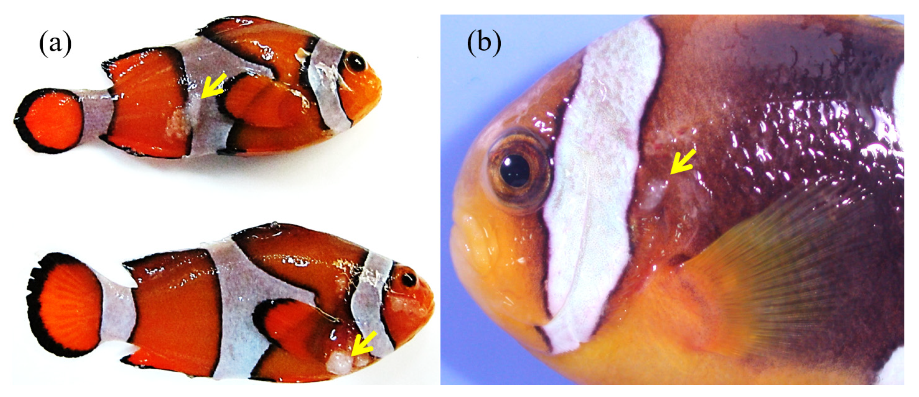
3.1. Macroscopic Observations

The fish samples showed sparse, multifocal, white, stiff, papilloma-like nodules on the body, skin, gills and fins in A. ocellaris. A. clarkii had only mild symptoms found on the operculum skin (Figure 1a,b).

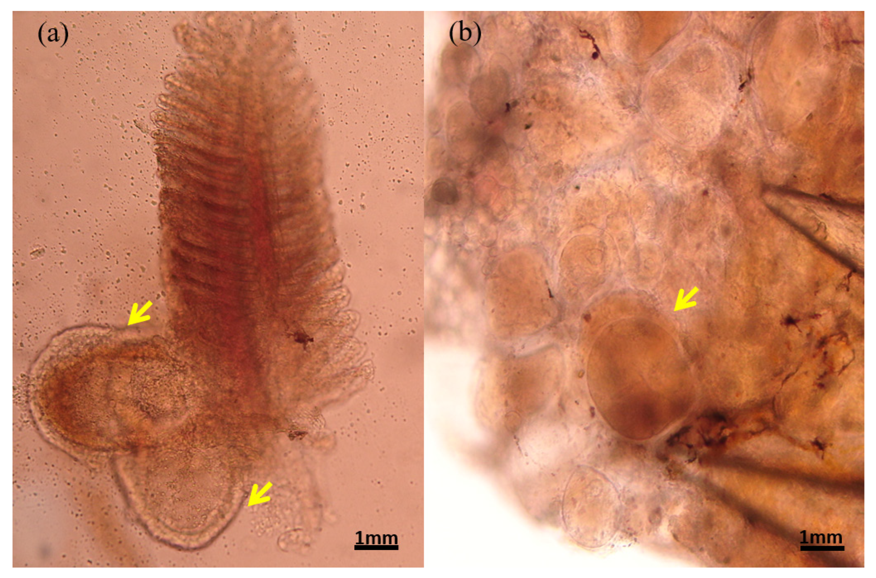
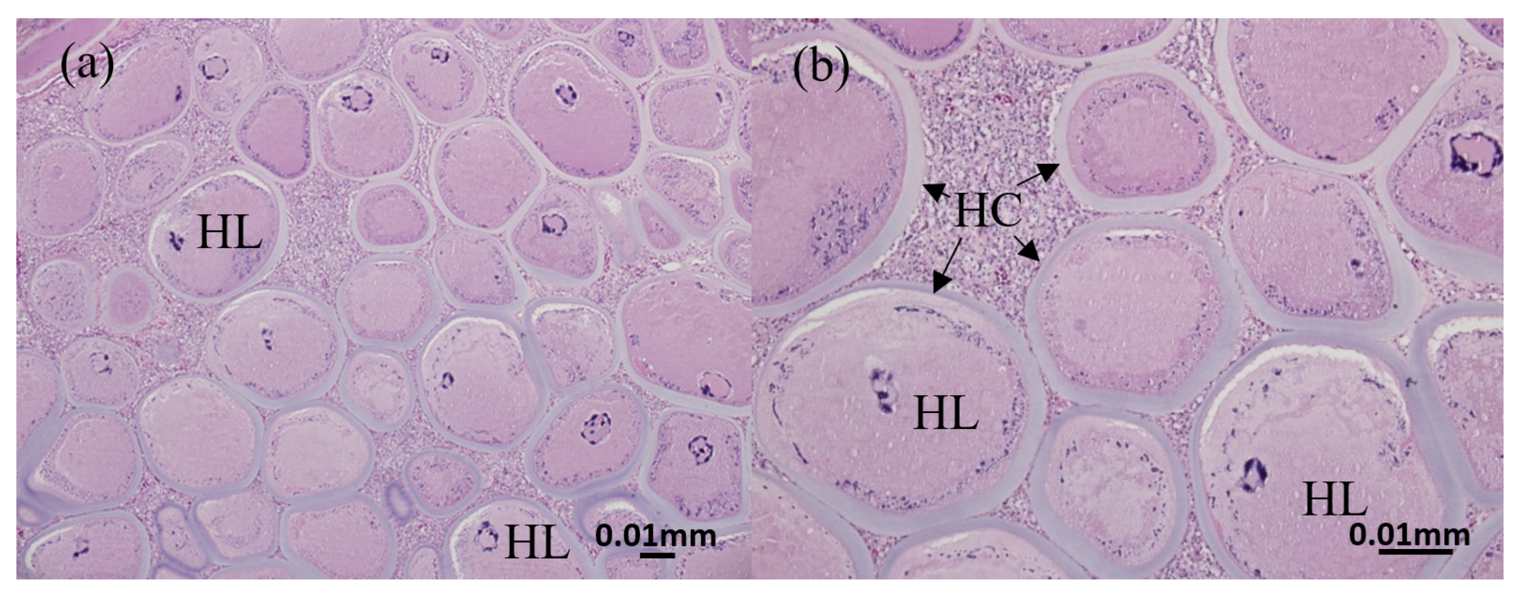
3.2. Histopathology of Nodules from Diseased Clownfish

The gill and fin are wreathed by an unusually thick outer membrane, forming a hyaline capsule (Figure 2a,b). The hyaline capsule is shown with a thick and smooth layer covering the hypertrophic lymphocystis cells (Figure 3a,b). The hypertrophic cells are present in various sizes. In addition, the magnified hypertrophic lymphocystis cells showed an irregular nucleus, and intracytoplasmic inclusion bodies around the cell membrane were strongly stained by hematoxylin and eosin (H&E) (Figure 4a,b).

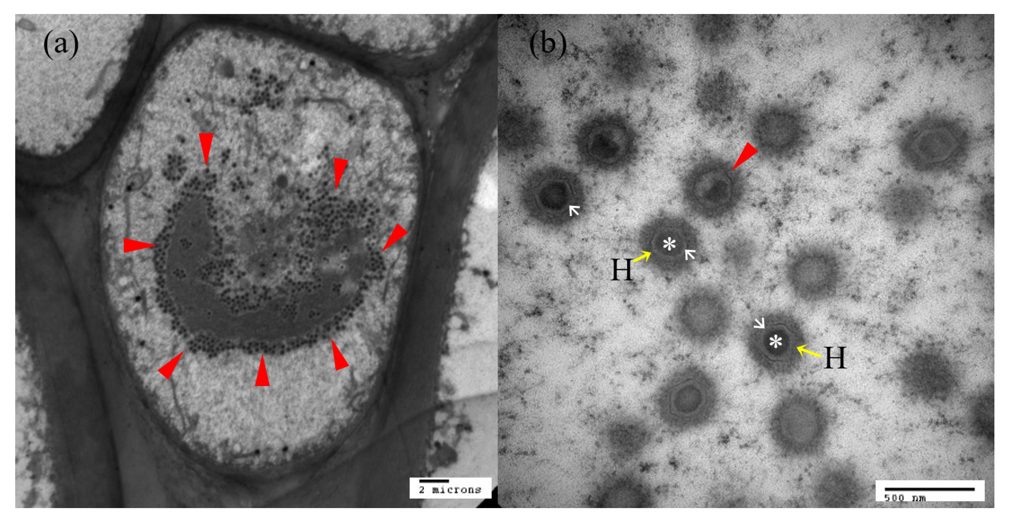
3.3. Electron Microscope

The existence of viral particles in the nodules of affected parts was observed by transmission electron micrographs (Figure 5a,b). Transmission electron microscopy (TEM) of the diseased fish shows large numbers of the virus particle (Figure 5a) in the cytoplasm of infected cells, with virions 180–230 nm in diameter, hexagonal in shape and with an inner dense nucleoid (Figure 5b).

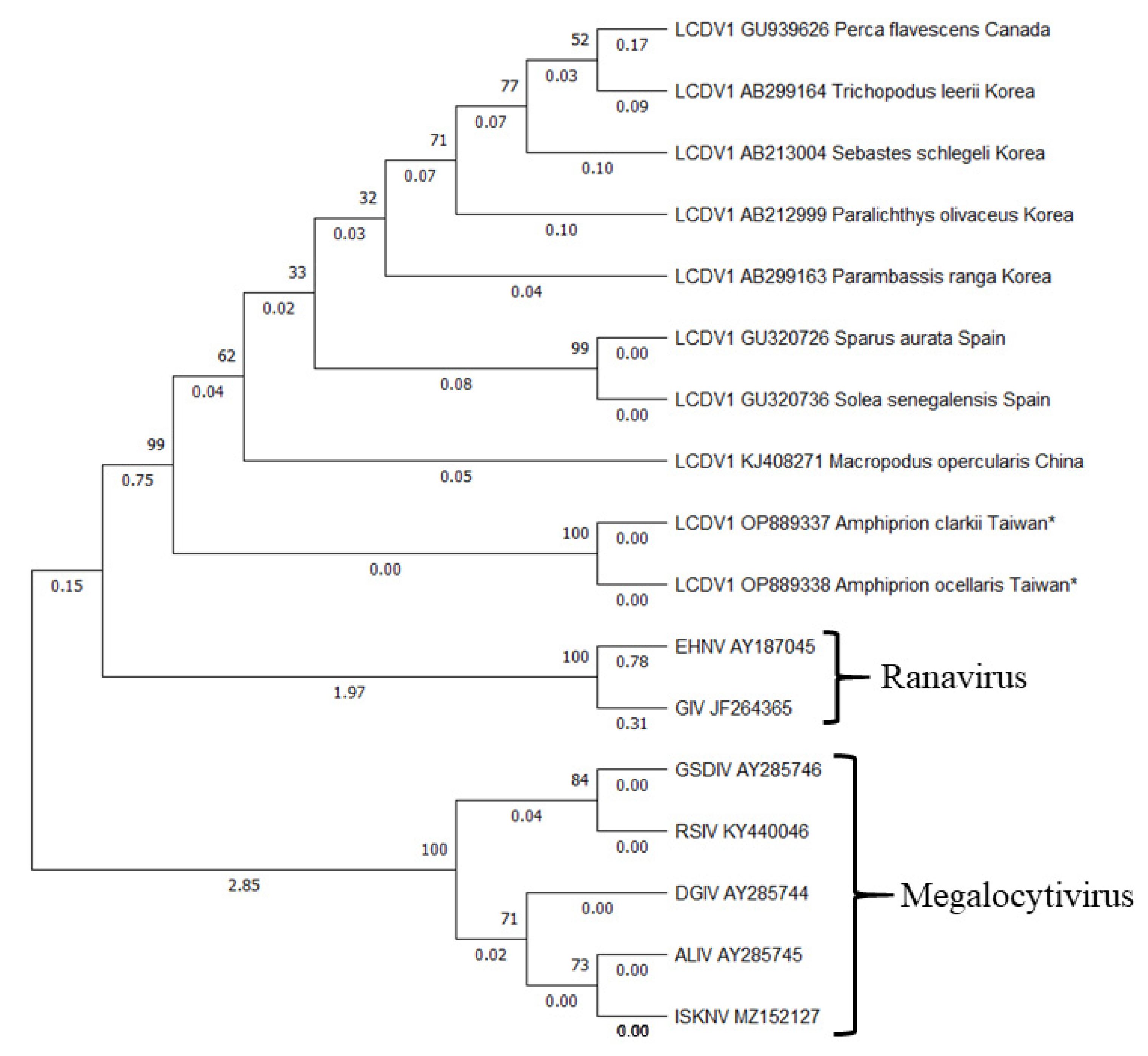
3.4. Molecular Phylogeny

Five clownfish, A. ocellaris (n = 4) and A. clarkii (n = 1), were detected with LCDV infection through a genetic study. The gene sequencing from the MCP of five fish showed positive results in gel electrophoresis and had a 1390 bp length in their PCR product. Three of the selected organs of A. ocellaris showed positive signs of LCDV infection; while A. clarkii had mild symptoms with positive results in the gill and fin; LCDV was not detected in the spleen (Table 2).
After aligning all the sequences from A. ocellaris (n = 4) and A. clarkii (n = 1), the gene sequences were grouped into two different groups: AOD100112 (A. clarkii) and AOD100090-2A (A. ocellaris). A phylogenetic tree based on the MCP gene sequences of these two genes was constructed by the maximum-likelihood method using Molecular Evolutionary Genetics Analysis (MEGA 11). To further ascertain the phylogenetic relationship between the new genotype LCDV and other iridovirus isolates, another four iridovirus core genes, MCP, DNA polymerase, myristoylated membrane protein (MMP) and ribonucleotide reductase (RNR) were amplified. Phylogenetic trees were constructed using MEGA 11 based on the MCP gene sequences of these genes. As shown in Figure 6, both of the LCDV virus genes in this study showed 100% similarity and grouped into the same cluster. Similar results were also found from the LCDV found in Spain (accession numbers GU320726 and GU320736) that had 99.7% similarity. The phylogenetic analysis consistently revealed that the LCDV Taiwan cluster (AOD100112 and AOD100090-2A) shared the closest relationship to LCDV found in China (accession number KJ408271) (pairwise distance: 92.5%), followed by Spain (accession number GU320726 and GU320736) (pairwise distance: 90.8–90.5%), Korea (accession number AB212999, AB213004, AB299163 and AB299164) (pairwise distance: 91.5–80.5%), Canada (accession number GU939626) (pairwise distance: 83%) and other iridovirus isolates (pairwise distance: 57.3–55.7%) (Table 3). Taken together, we proposed that G-VII was a novel LCDV isolate in the family Iridoviridae.

4. Discussion

A unique LCDV strain was discovered in captive-bred clownfish in Taiwan, representing the first gene sequencing of LCDV in clownfish. Although LCDV was known to infect a wide range of fish hosts [1,2], cases in Taiwan were still undiscovered. In Asia, LCDV was commonly reported in high-value aquaculture fishes: flounder, Paralichthys spp. [26,34,35] and rockfish Sebastes spp. [28,36,37]. In addition, LCDV infection is frequently detected in farmed gilt-head bream, Sparus aurata, in Egypt [38], Spain [27] and Tunisia [39]. As LCDV could infect a wide range of species of fish [1,2], clownfish may also become a victim. Before the study of Lam et al. [22], the detection of lymphocystic disease had only detected by histopathology and electron microscopy [7,21]. The reported LCDV infection in clownfish were found to be in A. ocellaris [7,21] and A. percula [21,22]. In histopathological studies, each white nodule consists of one or more lymphocystic cells with an irregular nucleus, enlarged cytoplasm and intracytoplasmic inclusion bodies surrounding the cell membrane, which corresponds to the previous research in clownfish [21,22]. Numerous viral particles and the distinct structure of the viral particle could be found under TEM. The LCDV viral particle from S. aurata [38], Solea senegalensis [40], Sebastes schlegelii [37] and A. ocellaris [7] have a similar ultrastructure morphology (hexagonal in shape with an inner dense nucleoid). In the present study, 9 species of clownfish (A. ocellaris, A. frenatus, A. latezonatus, Premnas biaculeatus, A. ocellaris var., A. percula, A. clarkii, A. ephippium and Amphiprion sp. cf. clarkii) were cultured in the same aquaculture system. However, only 8 fish from 2 species of clownfish, A. ocellaris (n = 7) and A. clarkii (n = 1), visually (white nodules on the external body structure: skin, fin and tail) and genetically showed LCDV infection. This is the first report demonstrating the presence of LCDV in A. clarkii. The infection was also confirmed by the degree of lesions and PCR. In recent studies, LCDV not only appears in the fin and skin of the fish host, but also internal organs such as the brain, liver, kidney, spleen and intestine [16,17,41]. The gene expression profile showed that the target organs of the LCDV are found to be the fin/skin, kidney, gut and liver [42,43,44,45]. In early infection, the LCDV enters the target organ from the bloodstream. Then, LCDV will spread to other organs during the virus outbreak or host immune suppression [42,43]. The present study showed both species of clownfish showed LCDV in the target organ (fin). However, LCDV was only found in the spleen of A. ocellaris, which indicates susceptibility to this virus is highest in A. ocellaris, followed by A. clarkii; the other species of clownfish are less sensitive. Moreover, the previous study showed the viral load was reduced progressively over the experiment infection time. The detection of the viral MCP gene transcription was detected in the kidney, intestine, skin-fin, liver and brain (except spleen) at 5 to 7 dpi, and only found in skin–caudal fin pools at 15 dpi [42]. This could be the reason for the mild symptoms of LCDV infection (no detection in the spleen) in A. clarkii which was sampled after 3 weeks of the LCDV outbreak.
Each genotype of LCDV is limited to a few or one species of fish, and infection of the same genotype of LCDV in different species of fish hosts is rare [12,13,28]. There is only one research study in Spain that found that LCDV genotype VII was able to cause infection in S. aurata (GU320726) and S. senegalensis (GU320736). The first detection of this LCDV genotype was found in cultured S. aurata and continued with infection in S. senegalensis after 3 months in the same fish farm. The LCDVs in genotype VII were found to have 99.7% similarity [12]. In the present study, the outbreak of LCDV in A. ocellaris and A. clarkii occurred in the same period, and the isolated MCP sequences of the two clownfish are 100% similar, representing the same iridovirus spreading in the system. This result could be similar to the infection of LCDV genotype VII, but the present study occurred in the same family of fish hosts and had different phenotypes of infection, even though the genetic relation of LCDV is more dependent on the species of host rather than geographical factors [28]. However, there is no full sequence of the MCP region of LCDV from clownfish in other research, and the ML polygenetic study showed that the genetic relationship of LCDV in the present research was related to geographical factors. The LCDV strains found in this study were closely related to the LCDV strains from paradise fish, Macropodus opercularis (KJ408271) (pairwise distance: 92.5%) from China, then followed by the strain from Spain (GU320726 and GU320736) (pairwise distance: 90.8–90.5%), Korea (AB299163, AB212999, AB213004 and AB299164) (pairwise distance: 91.5–80.5%) and lastly Canada (GU939626) (pairwise distance: 83%). Compared with the other 6 fish iridovirus genes, there was 57.3–55.7% similarity toward the isolated MCP sequence fen LCDV in the present study.

5. Conclusions

This is the first study that has provided new genotype molecular evidence of lymphocystis disease virus in clownfish, which might contribute greatly to the diagnosis and control of clownfish viral diseases. Although the vulnerability of fish to stress and disease could be enhanced by intensive breeding conditions, healthy management with good husbandry, early detection of disease pathogens and improved nutrition could eliminate disease outbreaks.

Author Contributions

Conceptualization, S.-C.C. and M.-A.T.; methodology, M.-C.C., P.-C.W. and M.-A.T.; software, M.-C.C. and M.S.S.; validation, M.-C.C., S.-C.C. and Y.-S.H.; formal analysis, M.-C.C., M.S.S. and S.-C.C.; investigation, M.-C.C., P.-C.W., Y.-S.H. and M.-A.T.; resources, S.-C.C. and Y.-S.H.; data curation, M.S.S. and Y.-T.K.; writing—original draft preparation, M.S.S., M.-C.C. and Y.-T.K.; writing—review and editing, M.S.S., M.-A.T. and S.-C.C.; visualization, M.S.S. and Y.-T.K.; supervision, S.-C.C. and M.-A.T.; project administration, S.-C.C. and M.-A.T.; funding acquisition, S.-C.C. All authors have read and agreed to the published version of the manuscript.

Funding

This research received no external funding.

Institutional Review Board Statement

The study was conducted according to the guidelines of the Animal Use Protocol and the Institutional Animal Care and Use Committee (IACUC) of the National Pingtung University of Science and Technology, Taiwan, under protocol number NPUST-108-042, 2019. The present study not involved experimental culturing, experimental infection and live sample collection. All of the animals were diseased before send for diagnosis (IACUC number: NPUST-108-042) in National Pingtung University of Science and technology. It considers that this type of project has no impact on animal welfare because all procedures are carried out after the diseased animals have been sacrificed as detection sample.

Informed Consent Statement

Not applicable.

Data Availability Statement

Not applicable.

Conflicts of Interest

The authors declare no conflict of interest.

References

  1. Anders, K. Lymphocystis disease of fishes. In Viruses of Lower Vertebrates; Ahne, W., Kurstak, E., Eds.; Springer: Berlin, Germany, 1989; pp. 140–160. [Google Scholar]
  2. Wolf, K. Fish Viruses and Fish Viral Disease; Cornell University Press: Ithaca, NY, USA, 1988. [Google Scholar]
  3. Peter, N.; Schmidt, W. Formation and disintegration of virion in lymphocystis cells of plaice Pleuronectes plassa. Dis. Aquat. Org. 1995, 21, 109–113. [Google Scholar] [CrossRef] [Green Version]
  4. Chinchar, V.G.; Essbauer, S.M.; He, J.G.; Hyatt, A.; Miyazaki, T.; Seligy, V.; Williams, T. Family Iridoviridae. In Virus Taxonomy: Eighth Report of the International Committee on Taxonomy of Viruses; Fauquet, C.M., Mayo, M.A., Maniloff, J., Desselberg, U., Ball, L.A., Eds.; Elsevier: Amsterdam, The Netherlands, 2005; pp. 145–162. [Google Scholar]
  5. Weissenberg, R. Studies on lymphocystis tumor cells of fish; the osmiophilic granules of the cytoplasmic inclusions and their interpretation as elementary bodies of the lymphocystis virus. Cancer Res. 1949, 9, 537–542. [Google Scholar] [PubMed]
  6. López-Bueno, A.; Mavian, C.; Labella, A.M.; Castro, D.; Borrego, J.J.; Alcami, A.; Alejoc, A. Concurrence of Iridovirus, Polyomavirus, and a unique member of a new group of fish papillomaviruses in lymphocystis disease-affected Gilthead Sea Bream. J Virol. 2016, 90, 8768–8779. [Google Scholar] [CrossRef] [PubMed] [Green Version]
  7. Pirarat, N.; Pratakpiriya, W.; Jongnimitpaiboon, K.; Sajjawiriyakul, K.; Rodkhum, C.; Chansue, N. Lymphocystis disease in cultured false clown anemonefish (Amphiprion ocellaris). Aquaculture 2011, 315, 414–416. [Google Scholar] [CrossRef]
  8. Xu, L.W.; Feng, J.; Huang, Y.H. Identification of lymphocystis disease virus from paradise fish Macropodus opercularis (LCDV-PF). Arch. Virol. 2014, 159, 2445–2449. [Google Scholar] [CrossRef]
  9. Tidona, C.A.; Darai, G. The complete DNA sequence of lymphocystis disease virus. Virology 1997, 230, 207–216. [Google Scholar] [CrossRef] [Green Version]
  10. Zhang, Q.Y.; Xiao, F.; Xie, J.; Li, Z.Q.; Gui, J.F. Complete genome sequence of lymphocystis disease virus isolated from China. J Virol. 2004, 78, 6982–6994. [Google Scholar] [CrossRef] [Green Version]
  11. Doszpoly, A.; Kaján, G.L.; Puentes, R.; Perretta, A. Complete genome sequence and analysis of a novel lymphocystivirusdetected in whitemouth croaker (Micropogonias furnieri): Lymphocystis disease virus 4. Arch. Virol. 2020, 165, 1215–1218. [Google Scholar] [CrossRef] [Green Version]
  12. Cano, I.; Valverde, E.J.; Lopez-Jimena, B.; Alonso, M.C.; Garcia-Rosado, E.; Sarasquete, C.; Borrego, J.J.; Castro, D. A new genotype of Lymphocystivirus isolated from cultured gilthead seabream, Sparus aurata L., and Senegalese sole, Solea senegalensis (Kaup). J. Fish Dis. 2010, 33, 695–700. [Google Scholar] [CrossRef]
  13. Palmer, L.J.; Hogan, N.S.; van den Heuvel, M.R. Phylogenetic analysis and molecular methods for the detection of lymphocystis disease virus from yellow perch, Perca flavescens (Mitchell). J. Fish Dis. 2012, 35, 661–670. [Google Scholar] [CrossRef]
  14. van den Heuvel, M.R.; Power, M.; MacKinnon, M.D.; Van Meer, T.; Dobson, E.P.; Dixon, D.G. Effects of oil sands related aquatic reclamation on yellow perch (Perca flavescens). I. Water quality characteristics and perch physiological and population responses. Can. J. Fish. Aquat. Sci. 1999, 56, 1213–1225. [Google Scholar] [CrossRef]
  15. Leiva-Rebollo, R.; Labella, A.M.; Valverde, E.J.; Castro, D.; Borrego, J.J. Persistence of Lymphocystis Disease Virus (LCDV) in Seawater. Food Environ. Virol. 2020, 12, 174–179. [Google Scholar] [CrossRef] [PubMed]
  16. Rahmati-Holasoo, H.; Ahmadivand, S.; Marandi, A.; Shokrpoor, S.; Palić, D.; Jahangard, A. Identification and characterization of lymphocystis disease virus (LCDV) from Indian glassy fish (Parambassis ranga Hamilton, 1822) in Iran. Aquac. Int. 2022, 30, 2593–2602. [Google Scholar] [CrossRef]
  17. Rahmati-Holasoo, H.; Ghalyanchilangeroudi, A.; Kafi, Z.Z.; Marandi, A.; Shokrpoor, S.; Imantalab, B.; Mousavi, H.E. Detection of lymphocystis disease virus (LCDV) from yellowbar angelfish (Pomacanthus maculosus Forsskål, 1775) in Iran: Histopathological and phylogenetic analysis. Aquaculture 2023, 562, 738862. [Google Scholar] [CrossRef]
  18. Wabnitz, C.; Taylor, M.; Green, E.; Razak, T. From Ocean to Aquarium; UNEP World Conservation Monitoring Centre: Cambridge, UK, 2003; p. 64. [Google Scholar]
  19. Abol-Munafi, A.B.; Borazmi-Lokman, N.H.; Asma, N.A.; Sarmiza, S.; Abduh, M.Y. Histological study on the gonad of the protadrous anemonefish (Amphiprion acellaris). J. Anim. Vet. Adv. 2011, 10, 3031–3036. [Google Scholar]
  20. Cato, J.C.; Brown, C.I. Marine Ornamental Collection, Culture, and Conservation. Open Fish Sci. J. 2003, 2, 55–58. [Google Scholar]
  21. Siva, U.; Marudhupandi, T.; Haq, M.A.B.; Thankappan, T.; Kumar, A. Histopathological study of lymphocystis disease virus (LCDV) in cultured false clownfish, Amphiprion ocellaris (Cuvier, 1830) and true clownfish, Amphiprion percula (Lacepede, 1802). J. Coast. Life Med. 2014, 2, 264–269. [Google Scholar]
  22. Lam, C.; Khairunissa, I.; Damayanti, L.; Kurobe, T.; Teh, S.J.; Pfahl, H.; Rapi, S.; Janetski, N.; Baxa, D.V. Detection of a new strain of lymphocystis disease virus (LCDV) in captive-bred clownfish Amphiprion percula in South Sulawesi, Indonesia. Aquac. Int. 2020, 28, 2121–2137. [Google Scholar] [CrossRef]
  23. Cheng, L.; Lin, W.H.; Wang, P.C.; Tsai, M.A.; Ho, P.Y.; Hsu, J.P.; Chen, R.S.; Chen, S.C. Epidemiology and phylogenetic analysis of Taura syndrome virus in cultured Pacific white shrimp Litopenaeus vannamei B. in Taiwan. Dis. Aquat. Org. 2011, 97, 17–23. [Google Scholar] [CrossRef] [Green Version]
  24. Hossain, M.; Song, J.; Kitamura, S.; Jung, S.; Oh, M. Phylogenetic analysis of lymphocystis disease virus from tropical ornamental fish species based on a major capsid protein gene. J. Fish Dis. 2008, 31, 473–479. [Google Scholar] [CrossRef]
  25. Tamura, K.; Stecher, G.; Kumar, S. MEGA11: Molecular Evolutionary Genetic Analysis Version 11. Mol. Biol. Evol. 2021, 38, 3022–3027. [Google Scholar] [CrossRef] [PubMed]
  26. Zan, J.D.; Sun, X.Q.; Zhang, Z.W.; Qu, L.Y.; Zhang, J.X. Application of quantitative PCR method in detection of Lymphocystis disease virus China (LCDV-cn) in Japanese flounder (Paralichthys olivaceus). Chin. J. Oceanol. Limnol. 2007, 25, 418–422. [Google Scholar] [CrossRef]
  27. Cano, I.; Alonso, M.C.; Garcia-Rasado, E.; Saint-Jean, S.R.; Castro, D.; Borrego, J.J. Detection of lymphocystis disease virus (LCDV) in asymptomatic cultured gilt-head seabream (Sparus aurata, L.) using an immunoblot technique. Vet. Microbiol. 2006, 113, 137–141. [Google Scholar] [CrossRef] [PubMed]
  28. Kitamura, S.I.; Jung, S.J.; Kim, W.S.; Nishizawa, T.; Yoshimizu, M.; Oh, M.J. A new genotype of lymphocystivirus, LCDV-RF, from lymphocystis disease rockfish. Arch. Virol. 2006, 151, 607–615. [Google Scholar] [CrossRef]
  29. Huang, S.M.; Tu, C.; Tseng, C.H.; Huang, C.C.; Kuo, H.C.; Chang, S.K. Genetic analysis of fish iridovirus isolated in Taiwan during 2001–2009. Arch. Virol. 2011, 156, 1505–1515. [Google Scholar] [CrossRef] [Green Version]
  30. Marsh, I.B.; Whittington, R.J.; O’Rourke, B.; Hyatt, A.D.; Chisholm, O. Rapid differentiation of Australian, European and American ranaviruses based on variation in major capsid protein gene sequence. Mol. Call. Probes. 2002, 16, 137–151. [Google Scholar] [CrossRef]
  31. Sudthongkong, C.; Miyata, M.; Miyazaki, T. Viral DNA sequences of genes encoding the ATPase and the major capsid protein of tropical iridovirus isolates which are pathogenic to fishes in Japan, South China Sea and Southeast Asian countries. Arch. Virol. 2002, 147, 2089–2109. [Google Scholar] [CrossRef]
  32. Dong, H.T.; Jitrakorn, S.; Kayansamruaj, P.; Pirarat, N.; Rodkhum, C.; Tattanarojpong, T.; Senapin, S.; Saksmerprome, V. Infectious spleen and kidney necrosis disease (ISKND) outbreaks in farmed barramundi (Lates calcarifer) in Vietnam. Fish Shellfish Immunol. 2017, 86, 65–73. [Google Scholar] [CrossRef]
  33. Swaminathan, T.R.; Raj, N.S.; Preena, P.G.; Pradhan, P.K.; Sood, N.; Kumar, R.G.; Sudhagar, A.; Sood, N.K. Infectious spleen and kidney necrosis virus-associated large-scale mortality in farmed giant gourami, Osphronemus goramy, in India. J. Fish Dis. 2021, 44, 2043–2053. [Google Scholar] [CrossRef]
  34. Kawato, S.; Nozaki, R.; Hirono, I.; Kondo, H. Genome Sequence of Lymphocystis Disease Virus 2 LCDV-JP_Oita_2018, Isolated from a Diseased Japanese Flounder (Paralichthys olivaceus) in Japan. Microbiol. Resour. Announc. 2021, 10, e00547-e21. [Google Scholar] [CrossRef]
  35. Zhan, W.B.; Li, Y.G.; Sheng, X.Z.; Xing, J.; Tang, X.Q. Detection of lymphocystis disease virus in Japanese flounder Paralichthys olivaceus and other marine teleosts from northern China. Chin. J. Oceanol. Limnol. 2010, 28, 1213–1220. [Google Scholar] [CrossRef]
  36. Sheng, X.Z.; Zheng, W.B.; Xu, S.J.; Cheng, S.F. Histopathological observation of lymphocystis disease and lymphocystis disease virus (LCDV) detection in cultured diseased Sebastes schlegeli. J. Ocean Univ. China 2007, 6, 378–382. [Google Scholar] [CrossRef]
  37. Zheng, F.R.; Liu, H.Z.; Guo, X.Y.; Wang, B. Isolation and identification of a new isolate of lymphocystis disease virus isolated from black rockfish (Sebastes schlegelii) in China. Aquaculture 2016, 451, 340–344. [Google Scholar] [CrossRef]
  38. Shawky, M.; Taha, E.; Ahmed, B.; Mahmoud, M.A.; Abdelaziz, M.; Faisal, M.; Yousif, A. Initial Evidence That Gilthead Seabream (Sparus aurata L.) Is a Host for Lymphocystis Disease Virus Genotype, I. Animals 2021, 11, 3032. [Google Scholar] [CrossRef]
  39. Hassad-Boubaker, S.; Bouzgarou, N.; Fakhfakh, E.; Khayech, M.; Mohamed, S.B.; Megdich, A.; Chéhida, N.B. Detection and genetic characterization of Lymphocystis Disease Virus (LCDV) isolated during disease outbreaks in cultured gilt-head sea bream Sparus aurata in Tunisia. Fish Pathol. 2013, 48, 101–104. [Google Scholar] [CrossRef] [Green Version]
  40. Alonso, M.C.; Cano, I.; Garcia-Rosado, E.; Cartro, D.; Lamas, J.; Berja, J.L.; Borrego, J.J. Isolation of lymphocystis disease virus from sole, Solea senegalensis Kaup, and blackspot sea bream, Pagellus bogaraveo (Brunnich). J. Fish Dis. 2005, 28, 221–228. [Google Scholar] [CrossRef]
  41. Kvitt, H.; Heinisch, G.; Diamant, A. Detection and phylogeny of Lymphocystivirus in sea bream Sparus aurata based on the DNA polymerase gene and major capsid protein sequences. Aquaculture 2008, 275, 58–63. [Google Scholar] [CrossRef]
  42. Carballo, C.; Castro, D.; Borrego, J.J.; Manchado, M. Gene expression profiles associated with lymphocystis disease virus (LCDV) in experimentally infected Senegalese sole (Solea senegalensis). Fish Shellfish Immunol. 2017, 66, 129–139. [Google Scholar] [CrossRef]
  43. Hu, G.B.; Lou, H.M.; Dong, X.Z.; Liu, Q.M.; Zhang, S.C. Characteristics of the interferon regulatory factor 5 (IRF5) and its expression in response to LCDV and poly I: C challenges in Japanese flounder, Paralichthys olivaceus. Dev. Comp. Immunol. 2012, 38, 377–382. [Google Scholar] [CrossRef]
  44. Valverde, E.J.; Borrego, J.J.; Sarasquete, M.C.; Ortiz-Delgado, J.B.; Castro, D. Target organs for lymphocystis disease virus replication in gilthead seabream (Sparus aurata). Vet. Res. 2017, 48, 21. [Google Scholar] [CrossRef] [Green Version]
  45. Sheng, X.Z.; Zeng, J.; Zhong, Y.; Tang, X.Q.; Xing, J.; Chi, H.; Zhan, W.B. Peripheral blood B-lymphocytes are involved in lymphocystis disease virus infection in flounder (Paralichthys olivaceus) via cellular receptor-mediated mechanism. Int. J. Mol. Sci. 2022, 23, 9225. [Google Scholar] [CrossRef] [PubMed]
Figure 1. Macroscopic view of Lymphocystis disease virus (LCDV) infected Amphiprion ocellaris (a) and A. clarkii (b). Yellow arrows are white nodules.
Animals 13 00153 g001
Figure 2. Macroscopic view of a 10× gill tissue slice (a) and a 10× fin tissue slice (b). Yellow arrow is a hyaline capsule.
Animals 13 00153 g002
Figure 3. Histopathological findings of hypertrophic lymphocystis cells. (a) 40× view and (b) 40× view. HL: hypertrophic lymphocystis cell; HC with arrow: hyaline capsule.
Animals 13 00153 g003
Figure 4. Histopathological findings of hypertrophic lymphocystis cells: (a) 100× view of single hypertrophic lymphocystic cell and (b) 100× view of lesion tissue on skin. HC: hyaline capsule; Arrowhead: intracytoplasmic inclusion bodies; N: nucleus.
Animals 13 00153 g004
Figure 5. Transmission electron microscopy image of an ultra-thin section of a hypertrophic lymphocystis cell. (a) Ultrastructure of a hypertrophic lymphocystis cell with an LCDV virus particle, (b) LCDV virus. Red arrow: virus particle; H with yellow arrow: hexagonal profile protein shell; White arrow: inner membrane; *: central DNA core.
Animals 13 00153 g005
Figure 6. Maximum likelihood polygenetic trees from Lymphocystivirus spp. based on the major capsid protein (MCP) gene under the K2+G (Kimura 2 + Gamma Distributed) model. Accession numbers were reported in the text. Number on node is 1000 bootstrap replications. * represent the present study.
Animals 13 00153 g006
Table 1. List of the MCP region sequences used in phylogenetic analysis.
Accession CodeVirus SpeciesHostRegionSizeReferences
OP889337LCDV 1Amphiprion clarkiiTaiwan1390Present study
OP889338LCDV 1Amphiprion ocellarisTaiwan1390Present study
AB299163LCDV 1Parambassis rangaKorea1337[26]
KJ408271LCDV 1Macropodus opercularisChina1208[8]
GU320726LCDV 1Sparus aurataSpain1317[12]
GU320736LCDV 1Solea senegalensisSpain1317[27]
AB212999LCDV 1Paralichthys olivaceusKorea1347[28]
GU939626LCDV 1Perca flavescensCanada1357[13]
AB299164LCDV 1Trichopodus leeriiKorea1337[24]
AB213004LCDV 1Sebastes schlegeliKorea1356[28]
JF264365GIVEpinephelus coioidesTaiwan1392[29]
AY187045EHNVPerca fluviatilisAustralia1472[30]
AY285746GSDIVEpinephelus chlorostigmaThailand1362[31]
KY440046RSIVLates calcariferVietnam1362[32]
AY285745ALIVAplocheilichthys centralisIndonesia1362[31]
MZ152127ISKNVOsphronemus goramyIndia1362[33]
AY285744DGIVColisa laliaMalaysia1362[31]
LCDV 1, Lymphocystis disease virus 1; GIV, Grouper iridovirus; EHNV, hematopoietic necrosis virus; GSDIV, Grouper sleepy disease iridovirus; RSIV, Red seabream iridovirus; ALIV, African lampeye iridovirus; ISKNV, Infectious spleen and kidney necrosis virus; DGIV, Dwarf gourami iridovirus.
Table 2. Detection of the Lymphocystis disease virus (LCDV) major capsid protein (MCP) gene from different organs of clownfish, Amphiprion spp.
Clownfish SpeciesOrgan
GillFinSpleen
Amphiprion clarkii (n = 1)++-
Amphiprion ocellaris (n = 4)+++
Table 3. Maximum likelihood polygenetic trees from Lymphocystivirus spp. based on the major capsid protein (MCP) gene under the K2+G (Kimura 2 + Gamma Distributed) model. Accession numbers were reported in the text. Number on node is 1000 bootstrap replications.
No.Accession Number12345678910111213141516
1OP889337/
OP889338
100.092.591.590.890.583.483.081.380.557.355.955.955.955.955.755.7
2KJ408271 100.089.989.689.286.083.883.983.356.652.854.454.254.254.154.2
3AB299163 100.089.188.886.182.984.385.255.752.254.253.753.553.554.1
4GU320726 100.099.784.883.483.284.056.153.654.854.154.154.154.7
5GU320736 100.084.583.182.983.755.953.654.754.054.054.054.5
6AB212999 100.082.384.885.656.350.852.251.951.751.652.2
7GU939626 10084.683.555.851.053.853.353.353.153.7
8AB299164 100.087.454.949.252.752.352.252.152.7
9AB213004 100.054.349.952.451.150.950.952.2
10GIV 100.069.950.750.750.950.550.5
11EHNV 100.054.654.554.554.354.7
12RSIV 100.095.094.694.999.4
13ALIV 100.099.799.794.7
14ISKNV 100.099.394.4
15DGIV 100.094.6
16GSDIV 100.0
Disclaimer/Publisher’s Note: The statements, opinions and data contained in all publications are solely those of the individual author(s) and contributor(s) and not of MDPI and/or the editor(s). MDPI and/or the editor(s) disclaim responsibility for any injury to people or property resulting from any ideas, methods, instructions or products referred to in the content.

