当前位置:
X-MOL 学术
›
Microsc. Res. Tech.
›
论文详情
Our official English website, www.x-mol.net, welcomes your feedback! (Note: you will need to create a separate account there.)
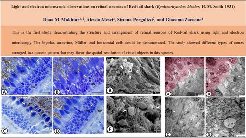
Light and electron microscopic observations on retinal neurons of red-tail shark (Epalzeorhynchos bicolor H. M. Smith, 1931)
Microscopy Research and Technique ( IF 2.5 ) Pub Date : 2024-01-08 , DOI: 10.1002/jemt.24488 Doaa M. Mokhtar 1, 2 , Alessio Alesci 3 , Simona Pergolizzi 3 , Giacomo Zaccone 4
Microscopy Research and Technique ( IF 2.5 ) Pub Date : 2024-01-08 , DOI: 10.1002/jemt.24488 Doaa M. Mokhtar 1, 2 , Alessio Alesci 3 , Simona Pergolizzi 3 , Giacomo Zaccone 4
Affiliation
|
The structure of photoreceptors (PR) and the arrangement of neurons in the retina of red-tail shark were investigated using light and electron microscopy. The PR showed a mosaic arrangement and included double cones, single cones (SC), and single rods. Most cones occur as SC. The ratio between the number of cones and rods was 3:1.39 (±0.29). The rods were tall that reached the pigmented epithelium. The outer plexiform layer (OPL) showed a complex synaptic connection between the horizontal and photoreceptor terminals that were surrounded by Müller cell processes. Electron microscopy showed that the OPL possessed both cone pedicles and rod spherules. Each rod spherule consisted of a single synaptic ribbon within the invaginating terminal endings of the horizontal cell (hc) processes. In contrast, the cone pedicles possessed many synaptic ribbons within their junctional complexes. The inner nuclear layer consisted of bipolar, amacrine, Müller cells, and hc. Müller cells possessed intermediate filaments and cell processes that can reach the outer limiting membrane and form connections with each other by desmosomes. The ganglion cells were large multipolar cells with a spherical nucleus and Nissl’ bodies in their cytoplasm. The presence of different types of cones arranged in a mosaic pattern in the retina of this species favors the spatial resolution of visual objects.
中文翻译:

对红尾鲨视网膜神经元的光和电子显微镜观察(Epalzeorhynchos bicolor HM Smith,1931)
使用光学和电子显微镜研究了红尾鲨视网膜中光感受器(PR)的结构和神经元的排列。 PR 呈马赛克排列,包括双锥体、单锥体 (SC) 和单杆体。大多数视锥细胞以 SC 形式出现。视锥细胞和视杆细胞的数量之比为3:1.39 (±0.29)。杆状细胞很高,可以到达色素上皮。外丛状层(OPL)显示水平终端和光感受器终端之间存在复杂的突触连接,这些终端被穆勒细胞突包围。电子显微镜显示 OPL 具有锥蒂和杆状球体。每个杆状小球由水平细胞(hc)突的内陷末端内的单个突触带组成。相比之下,锥蒂在其连接复合体中拥有许多突触带。内核层由双极细胞、无长突细胞、穆勒细胞和 hc 组成。穆勒细胞拥有中间丝和细胞突起,可以到达外界膜并通过桥粒相互形成连接。神经节细胞是大型多极细胞,其细胞质中具有球形核和尼氏体。该物种视网膜中存在以马赛克图案排列的不同类型的视锥细胞,有利于视觉物体的空间分辨率。
更新日期:2024-01-08
中文翻译:
对红尾鲨视网膜神经元的光和电子显微镜观察(Epalzeorhynchos bicolor HM Smith,1931)
使用光学和电子显微镜研究了红尾鲨视网膜中光感受器(PR)的结构和神经元的排列。 PR 呈马赛克排列,包括双锥体、单锥体 (SC) 和单杆体。大多数视锥细胞以 SC 形式出现。视锥细胞和视杆细胞的数量之比为3:1.39 (±0.29)。杆状细胞很高,可以到达色素上皮。外丛状层(OPL)显示水平终端和光感受器终端之间存在复杂的突触连接,这些终端被穆勒细胞突包围。电子显微镜显示 OPL 具有锥蒂和杆状球体。每个杆状小球由水平细胞(hc)突的内陷末端内的单个突触带组成。相比之下,锥蒂在其连接复合体中拥有许多突触带。内核层由双极细胞、无长突细胞、穆勒细胞和 hc 组成。穆勒细胞拥有中间丝和细胞突起,可以到达外界膜并通过桥粒相互形成连接。神经节细胞是大型多极细胞,其细胞质中具有球形核和尼氏体。该物种视网膜中存在以马赛克图案排列的不同类型的视锥细胞,有利于视觉物体的空间分辨率。


 京公网安备 11010802027423号